- Record: found
- Abstract: found
- Article: found
Endovascular treatment of an aortic aneurysm and patent ductus arteriosus
case-report
Read this article at
There is no author summary for this article yet. Authors can add summaries to their articles on ScienceOpen to make them more accessible to a non-specialist audience.
Abstract
Introduction
Aortic coarctation may occasionally be associated with a second congenital disease,
such as patent ductus arteriosus (PDA) (1). When diseases coexist in children or teenagers,
surgical treatment is frequently recommended as far as both conditions can be addressed
in a single surgery. Depending on the techniques used during the initial surgery,
late complications after surgery for aortic coarctation may be the formation of an
aneurysm in the descending aorta (2). Reopening of PDA has also been observed after
surgical ligation (3). Re-do surgery in these cases may prove challenging in elderly
patients, and endovascular techniques may be used for lower morbidity and mortality
risk (4).
Case Report
A 63-year-old man presented with progressive cough and dyspnea on exertion after a
large thoracic aneurysm in the post-isthmic aorta (TAA) was diagnosed on plain chest
X-ray. His symptoms were first attributed to his life-long smoking habit. He had undergone
surgery 35 years ago for aortic coarctation with dacron patch aortoplasty; the initial
surgical protocol did not mention ligation of PDA. Angio-computed tomography (angio-CT)
confirmed the presence of an 80-mm thoracic aortic aneurysm and a 7-mm PDA (arrow
in Fig. 1). The ascending aorta was moderately dilated and the aortic valve was tricuspid
and competent. The thoracic aorta distal to the aneurysm was significantly dilated
down to the diaphragm. The left subclavian artery was dilated at the origin. Echocardiography
showed normal systolic left ventricular function, moderate pulmonary hypertension
(sPAP=50 mm Hg), and confirmed significant left-to-right shunting (Qp/Qs=2.1). Coronary
angiography found no flow-limiting stenosis.
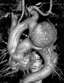
Figure 1
Diagnostic angio-CT. A large saccular aneurysm of the thoracic aorta and PDA can be
observed. The distal thoracic aorta is also enlarged
Endovascular treatment for both TAA and PDA was planned. To avoid potential type II
endoleaks, the patient underwent surgical ligation of the left subclavian artery and
left carotid–subclavian by-pass. Subsequently, he was brought intubated from the OR
to the cathlab, where a dedicated Occlutech device (Occlutech® Duct Occluder, Occlutech,
Helsingborg, Sweden) to close PDA was first deployed via the right common femoral
vein (Video 1, 2, 3 and 4). Two endovascular Valiant Captivia grafts (EVG) (Medtronic,
Santa Rosa, CA, USA) were then implanted distal to the ostium of the left carotid
artery across the ostium of left subclavian artery with complete exclusion of TAA
(Fig. 2, Video 5, 6 and 7). Cerebrospinal fluid (CSF) was repeatedly aspirated for
48 h because of CSF hypertension to avoid the risk of paraplegia. He made a full recovery
and was discharged on day 5 after endovascular treatment. The 3-year angio-CT follow-up
confirmed complete sealing of the aneurysm with a minor type 2 endoleak through an
intercostal artery, with no increase in the diameter of the aneurysm.
Video 1
Video 2
Video 3
Video 4
Figure 2
One-month follow-up angio-CT. The final result of the hybrid procedure shows a completely
excluded aortic aneurysm, surgically interrupted left subclavian artery, and completely
occluded PDA
Video 5
Video 6
Video 7
Discussion
Late complications after surgical repair of aortic coarctation may include aneurysm
formation in the post-isthmic thoracic aorta. They are rare after subclavian flap
aortoplasty, but are more common after dacron patch aortoplasty (2), such as that
observed in our case. Aneurysm formation carries the risk of rupture and sudden death,
whereas re-do surgery leads to a 14% in-hospital death rate or morbidity because of
paraplegia or bleeding (4). Small series of this high-risk surgical group of patients
have been successfully treated by endovascular stent-graft placement with lower peri-procedural
morbidity or mortality (4).
Meanwhile, PDA with a non-dilated descending aorta was treated with EVG insertion
in elderly patients to avoid the risk of rupture of a calcified duct on percutaneous
intervention (5), with good short-term result and no residual endoleaks (6). Some
reports describe percutaneous PDA closure with Amplatzer devices in patients with
thoracic aortic aneurysms leaving the latter untreated (7). Occasional reports mention
exclusion of both thoracic aortic aneurysm and PDA with stent-graft insertion in the
aorta (8), sometimes using open chest surgical techniques (9).
Considering the high risk of surgical complications in our patient, we decided to
treat TAA and PDA using endovascular techniques. Isolated EVAR was not considered
an option in our case because of pulmonary hypertension due to a large 7 mm PDA; closure
of PDA was considered necessary to avoid type II endoleaks from the aneurysm. For
the same reason, the left subclavian artery was ligated.
Covering of the whole descending thoracic aorta and exclusion of the left subclavian
artery by EVAR frequently leads to CSF hypertension, which is associated with a risk
of spinal cord ischemia and paraplegia (10). Close monitoring of CSF pressure and
continuous drainage, as necessary, may mitigate this risk, similar to that observed
in our case.
Conclusion
We present the case of a patient with late thoracic aneurysm formation after aortic
coarctation surgery associated with PDA. Endovascular treatment was successfully performed
immediately after surgical left subclavian debranching. The 3-year angio-CT follow-up
showed persistent optimal result of endovascular exclusion in both conditions.
Video 1
Diagnostic angio
Video 2
Snared wire in PA
Video 3
PDA occluder deployment
Video 4
PDA occluder deployed
Video 5
First Valiant EVG deployment
Video 6
Second Valiant Captivia deployed
Video 7
Final angiographic result.
Related collections
Most cited references10
- Record: found
- Abstract: not found
- Article: not found
Fate of patients with spinal cord ischemia complicating thoracic endovascular aortic repair
Kenneth DeSart, Salvatore T Scali, Robert Feezor … (2013)
- Record: found
- Abstract: found
- Article: not found
Percutaneous endovascular repair of aneurysm after previous coarctation surgery.
Bette Korber, Christoph Nienaber, Michael Petzsch … (2003)
- Record: found
- Abstract: found
- Article: not found
Open stent-grafting for adult patent ductus arteriosus with a distal aortic arch aneurysm.
Ben Sasaki, Kazuteru Shimizu, Nobuhisa Ohno … (2011)