Inviting an author to review:
Find an author and click ‘Invite to review selected article’ near their name.
Search for authorsSearch for similar articles
0
views
0
recommends
+1 Recommend
0 collections
    0
    shares
      • Record: found
      • Abstract: found
      • Article: found
      Is Open Access

      Morphology of the Esophageal Hiatus: Is It Different in 3 Types of Hiatus Hernias?

      research-article

      Read this article at

      Bookmark
          There is no author summary for this article yet. Authors can add summaries to their articles on ScienceOpen to make them more accessible to a non-specialist audience.

          Abstract

          Background/Aims

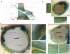
          The esophageal hiatus is formed by the right crus of the diaphragm in the majority of subjects. Contraction of the hiatus exerts a sphincter-like action on the lower esophageal sphincter (LES). The aim is to study the hiatal anatomy (using CT scan imaging) and function (using high-resolution manometry [HRM]), and esophageal motor function in patients with sliding and paraesophageal hiatal hernia.

          Methods

          We assessed normal subjects (n = 20), patients with sliding type 1 hernia (n = 18), paraesophageal type 2 hernia (n = 19), and mixed type 3 hernia (n = 19). Hernia diagnosis was confirmed on the upper gastrointestinal series. The hiatal morphology was constructed from the CT scan images. The LES pressure and relaxation, percent peristalsis, bolus pressure, and hiatal squeeze pressure were assessed by HRM.

          Results

          The CT images revealed that the esophageal hiatus is formed by the right crus of the diaphragm in all normal subjects and 86% of hernia patients. The hiatus is elliptical in shape with a surface area of 1037 mm 2 in normal subjects. The hiatal dimensions were larger in patients compared to normal subjects. The HRM revealed impaired LES relaxation and higher bolus pressure in patients with paraesophageal compared to the sliding hernia. The hiatal pinch on HRM was recognized in significantly higher number of patients with sliding as compared to paraesophageal hernia.

          Conclusions

          Using a novel approach, we provide details of the esophageal hiatus in patients with various kinds of hiatal hernia. Impaired LES relaxation in paraesophageal hernia may play a role in its pathophysiology and genesis of symptoms.

          Related collections

          Most cited references18

          • Record: found
          • Abstract: not found
          • Article: not found

          The esophagogastric junction.

            Bookmark
            • Record: found
            • Abstract: found
            • Article: found
            Is Open Access

            Preoperative diagnosis of hiatal hernia: barium swallow X-ray, high-resolution manometry, or endoscopy?

            Summary Background The assessment of hiatal hernias (HH) is typically done with barium swallow X‑ray, upper endoscopy, and by high-resolution esophageal manometry (HRM). The aim of this study was to assess the clinical utility of these methods in terms of HH detection and their correlation to gastroesophageal reflux disease (GERD). Methods A retrospective comparative analysis of patients with symptoms of GERD was carried out. The performance of endoscopy and HRM in diagnosing HH was assessed, taking barium swallow X‑ray as a reference. Furthermore, statistically comparative analysis between detected hernias and the presence of reflux disease in ambulatory impedance-pH monitoring (MII) was performed. Results Overall, 112 patients were analyzed. Barium swallow X‑ray showed no correlation either to HR manometrically or to endoscopically assessed HH. Significant accordance in the detection rate of HH was proved between HRM and gastroesophagoscopy (p < 0.001). Only endoscopically assessed HH showed a significant correlation with GERD (p = 0.047). No correlation between detected hernias and GERD could be found either with HRM or with barium swallow X‑ray. Conclusions Barium swallow X‑ray provided the highest rate of HH detection (76.8%). For the reliable exclusion of HH prior to treatment, all three mentioned investigations appear to be necessary in order of low conformity.
              Bookmark
              • Record: found
              • Abstract: found
              • Article: found
              Is Open Access

              Three-Dimensional Myoarchitecture of the Lower Esophageal Sphincter and Esophageal Hiatus Using Optical Sectioning Microscopy

              Studies to date have failed to reveal the anatomical counterpart of the lower esophageal sphincter (LES). We assessed the LES and esophageal hiatus morphology using a block containing the human LES and crural diaphragm, serially sectioned at 50 μm intervals and imaged at 8.2 μm/pixel resolution. A 3D reconstruction of the tissue block was reconstructed in which each of the 652 cross sectional images were also segmented to identify the boundaries of longitudinal (LM) and circular muscle (CM) layers. The CM fascicles on the ventral surface of LES are arranged in a helical/spiral fashion. On the other hand, the CM fascicles from the two sides cross midline on dorsal surface and continue as sling/oblique muscle on the stomach. Some of the LM fascicles of the esophagus leave the esophagus to enter into the crural diaphragm and the remainder terminate into the sling fibers of the stomach. The muscle fascicles of the right crus of diaphragm which form the esophageal hiatus are arranged like a “noose” around the esophagus. We propose that circumferential squeeze of the LES and crural diaphragm is generated by a unique myo-architectural design, each of which forms a “noose” around the esophagus.
                Bookmark

                Author and article information

                Journal
                J Neurogastroenterol Motil
                J Neurogastroenterol Motil
                Journal of Neurogastroenterology and Motility
                Korean Society of Neurogastroenterology and Motility
                2093-0879
                2093-0887
                January 2020
                30 January 2020
                : 26
                : 1
                : 51-60
                Affiliations
                [1 ]Department of Gastroenterology, University of California San Diego, La Jolla, CA, USA
                [2 ]Department of Radiology, University of California San Diego, La Jolla, CA, USA
                [3 ]Department of Surgery, University of California San Diego, La Jolla, CA, USA
                Author notes
                [* ]Correspondence: Ravinder K Mittal, MD, University of California San Diego, Altman Clinical and Translational Research Institute Building, 9500 Gillman Drive, MC 0061, La Jolla, CA 92093-0990, USA, Tel: +1-858-543-3328, E-mail: rmittal@ 123456ucsd.edu
                Article
                jnm-26-051
                10.5056/jnm18208
                6955188
                31677612
                ad3952dc-2f84-4657-9093-2edc8832bcfd
                © 2020 The Korean Society of Neurogastroenterology and Motility

                This is an Open Access article distributed under the terms of the Creative Commons Attribution Non-Commercial License ( http://creativecommons.org/licenses/by-nc/4.0) which permits unrestricted non-commercial use, distribution, and reproduction in any medium, provided the original work is properly cited.

                History
                : 18 December 2018
                : 04 July 2019
                : 23 July 2019
                Categories
                Original Article

                Neurology
                esophageal peristalsis,hiatal,hernia,lower esophageal sphincter,manometry,tomography x-ray computed

                Comments

                Comment on this article