12
views
0
recommends
+1 Recommend
1 collections
    0
    shares
      • Record: found
      • Abstract: found
      • Article: found
      Is Open Access

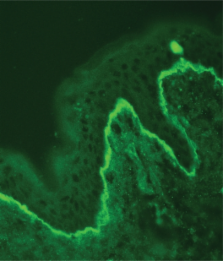
      The lupus band test in systemic lupus erythematosus patients

      review-article

      Read this article at

      ScienceOpenPublisherPMC
      Bookmark
          There is no author summary for this article yet. Authors can add summaries to their articles on ScienceOpen to make them more accessible to a non-specialist audience.

          Abstract

          The lupus band test (LBT) is a diagnostic procedure that is used to detect deposits of immunoglobulins and complement components along the dermoepidermal junction in patients with lupus erythematosus (LE). The LBT is positive in about 70%–80% of sun-exposed non-lesional skin specimens obtained from patients with systemic LE (SLE), and in about 55% of SLE cases if sun-protected nonlesional skin is analyzed. In patients with cutaneous LE only, the lesional skin usually shows a positive LBT. The LBT helps in differentiating LE from other similar skin conditions and may also be helpful in making the diagnosis of SLE in subjects with no specific cutaneous lesions. Furthermore, a positive LBT may be applied as a prognostic parameter for LE patients. However, the correct interpretation of this test requires detailed knowledge of the site of the biopsy, deposit components, morphology and brightness of the immunofluorescent band, and other associated serologic findings, as well as the response to treatment. It must be emphasized that LBT is a laboratory procedure that should always be interpreted in conjunction with clinical findings and other serological and immunopathological parameters.

          Most cited references20

          • Record: found
          • Abstract: found
          • Article: not found

          Incidence of systemic lupus erythematosus. Race and gender differences.

          To examine racial differences in the incidence of systemic lupus erythematosus (SLE). A population-based registry of SLE patients in Allegheny County, Pennsylvania, was used to identify incident cases of SLE diagnosed between January 1, 1985 and December 31, 1990, from 3 sources, by medical record review (University of Pittsburgh Lupus Databank, rheumatologists, and hospitals). Capture-recapture methods using log-linear models were used to estimate the level of case-finding and to calculate 95% confidence intervals (CI). Incidence rates were calculated per 100,000 population. A total of 191 definite and 78 probable incident cases of SLE were identified, and the overall annual incidence rates were 2.4 (95% CI 2.1-2.8) and 1.0 (95% CI 0.8-1.3), respectively. The crude incidence rates of definite SLE were 0.4 for white males, 3.5 for white females, 0.7 for African-American males, and 9.2 for African-American females. The annual incidence rates of definite SLE remained fairly constant over the study interval. African-American females with definite SLE had a younger mean age at diagnosis compared with white females (P < 0.05). Since the overall ascertainment rate was high (85%; 95% CI 78-92%), the ascertainment-corrected incidence rate for definite SLE, 2.8 (95% CI 2.6-3.2), was similar to the crude rate. Our rates clearly confirm previous reports of an excess incidence of SLE among females compared with males and among African-Americans compared with whites. We have used capture-recapture methods to improve the accuracy of SLE incidence rates, and we advocate their use to facilitate comparisons across studies.
            Bookmark
            • Record: found
            • Abstract: found
            • Article: not found

            The cutaneous pathology of lupus erythematosus: a review.

            The presentation of lupus erythematosus (LE) ranges from a skin rash unaccompanied by extracutaneous stigmata to a rapidly progressive lethal multiorgan disease. The diagnosis and subclassification is traditionally based on the correlation of serological and clinical findings. The latter include a photoinduced skin rash, arthralgia, arthritis, fever, Raynaud's phenomenon, anemia, leukopenia, serositis, nephritis and central nervous sysdtem disease. The conventional classification scheme includes systemic, subacute cutaneous and discoid LE. Recent advances in our understanding of the cutaneous histopathology which correlates with the traditional forms of LE, along with certain novel LE subtypes, are the focus of this review. In addition to the main subtypes of LE, we will discuss associated vasculopathic lesions and the contribution of immunofluorescence microscopy to the diagnosis of LE and related connective tissue disease syndromes. Consideration will be given to unusual variants of LE such as anti-Ro/SSA-positive systemic lupus erythematosus (SLE), bullous SLE, lymphomatoid LE, lupus erythematosus profundus, drug induced LE, linear cutaneous LE, chiblains LE and parvovirus B19-associated LE.
              Bookmark
              • Record: found
              • Abstract: found
              • Article: not found

              Survival study by organ disorders in 306 Japanese patients with systemic lupus erythematosus: results from a single center.

              Survival rate and causes of death according to the period of diagnosis and four accompanying organ disorders were analyzed in 306 Japanese patients with systemic lupus erythematosus. The survival rate was gradually improved, and the survival rate during 5- and 10-year periods of the patients diagnosed in 1990-2004 was 94 and 92%, 20-year period of those in 1980-1989 was 77%, 30-year period of those in 1975-1979 was 71%, respectively. Survival rate of those with serositis, pulmonary hypertension, and positive family history tended to be reduced, while that of the cases with neuropsychiatric disorder and renal disorder was significantly reduced. Overlapping of these organ disorders was an important factor for a poor prognosis. Bronchopneumonia and cerebrovascular accidents were frequent causes of death, and treatment for anti-phospholipid antibody syndrome and life-style diseases such as hypertension and arteriosclerosis was thought to be important for a good outcome.
                Bookmark

                Author and article information

                Journal
                Ther Clin Risk Manag
                Therapeutics and Clinical Risk Management
                Therapeutics and Clinical Risk Management
                Dove Medical Press
                1176-6336
                1178-203X
                2011
                2011
                24 January 2011
                : 7
                : 27-32
                Affiliations
                Department of Dermatology, Venereology, and Allergology, Wroclaw Medical University, Wroclaw, Poland
                Author notes
                Correspondence: Adam Reich, Department of Dermatology, Venereology, and Allergology, Wroclaw, Medical University, Ul. Chalubinskiego 1, 50-368 Wroclaw, Poland, Tel +48 605076722, Fax +48 713270942, Email adi_medicalis@ 123456go2.pl
                Article
                tcrm-7-027
                10.2147/TCRM.S10145
                3039011
                21339940
                ae583f25-2f0e-43b4-a2cb-aa37aa745967
                © 2011 Reich et al, publisher and licensee Dove Medical Press Ltd.

                This is an Open Access article which permits unrestricted noncommercial use, provided the original work is properly cited.

                History
                : 21 January 2011
                Categories
                Review

                Medicine
                dermoepidermal junction,lupus erythematosus,diagnostics
                Medicine
                dermoepidermal junction, lupus erythematosus, diagnostics

                Comments

                Comment on this article