0
views
0
recommends
+1 Recommend
0 collections
    0
    shares
      • Record: found
      • Abstract: found
      • Article: found
      Is Open Access

      Emotion processing in youths with conduct problems: an fMRI meta-analysis

      research-article

      Read this article at

      Bookmark
          There is no author summary for this article yet. Authors can add summaries to their articles on ScienceOpen to make them more accessible to a non-specialist audience.

          Abstract

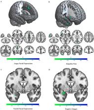
          Functional magnetic resonance imaging (fMRI) studies consistently indicate differences in emotion processing in youth with conduct problems. However, no prior meta-analysis has investigated emotion-specific responses associated with conduct problems. This meta-analysis aimed to generate an up-to-date assessment of socio-affective neural responding among youths with conduct problems. A systematic literature search was conducted in youths (ages 10–21) with conduct problems. Task-specific seed-based d mapping analyses examined responses to threatening images, fearful and angry facial expressions, and empathic pain stimuli from 23 fMRI studies, which included 606 youths with conduct problems and 459 comparison youths. Whole-brain analyses revealed youths with conduct problems relative to typically developing youths, when viewing angry facial expressions, had reduced activity in left supplementary motor area and superior frontal gyrus. Additional region of interest analyses of responses to negative images and fearful facial expressions showed reduced activation in right amygdala across youths with conduct problems. Youths with callous-unemotional traits also exhibited reduced activation in left fusiform gyrus, superior parietal gyrus, and middle temporal gyrus when viewing fearful facial expressions. Consistent with the behavioral profile of conduct problems, these findings suggest the most consistent dysfunction is found in regions associated with empathic responding and social learning, including the amygdala and temporal cortex. Youth with callous-unemotional traits also show reduced activation in the fusiform gyrus, consistent with reduced attention or facial processing. These findings highlight the potential role of empathic responding, social learning, and facial processing along with the associated brain regions as potential targets for interventions.

          Related collections

          Most cited references91

          • Record: found
          • Abstract: not found
          • Book: not found

          Diagnostic and Statistical Manual of Mental Disorders

            Bookmark
            • Record: found
            • Abstract: found
            • Article: not found

            Automated anatomical labeling of activations in SPM using a macroscopic anatomical parcellation of the MNI MRI single-subject brain.

            An anatomical parcellation of the spatially normalized single-subject high-resolution T1 volume provided by the Montreal Neurological Institute (MNI) (D. L. Collins et al., 1998, Trans. Med. Imag. 17, 463-468) was performed. The MNI single-subject main sulci were first delineated and further used as landmarks for the 3D definition of 45 anatomical volumes of interest (AVOI) in each hemisphere. This procedure was performed using a dedicated software which allowed a 3D following of the sulci course on the edited brain. Regions of interest were then drawn manually with the same software every 2 mm on the axial slices of the high-resolution MNI single subject. The 90 AVOI were reconstructed and assigned a label. Using this parcellation method, three procedures to perform the automated anatomical labeling of functional studies are proposed: (1) labeling of an extremum defined by a set of coordinates, (2) percentage of voxels belonging to each of the AVOI intersected by a sphere centered by a set of coordinates, and (3) percentage of voxels belonging to each of the AVOI intersected by an activated cluster. An interface with the Statistical Parametric Mapping package (SPM, J. Ashburner and K. J. Friston, 1999, Hum. Brain Mapp. 7, 254-266) is provided as a freeware to researchers of the neuroimaging community. We believe that this tool is an improvement for the macroscopical labeling of activated area compared to labeling assessed using the Talairach atlas brain in which deformations are well known. However, this tool does not alleviate the need for more sophisticated labeling strategies based on anatomical or cytoarchitectonic probabilistic maps.
              Bookmark
              • Record: found
              • Abstract: not found
              • Book: not found

              Multiple Imputation for Nonresponse in Surveys

                Bookmark

                Author and article information

                Contributors
                ka741@georgetown.edu
                Journal
                Transl Psychiatry
                Transl Psychiatry
                Translational Psychiatry
                Nature Publishing Group UK (London )
                2158-3188
                30 March 2023
                30 March 2023
                2023
                : 13
                : 105
                Affiliations
                [1 ]GRID grid.213910.8, ISNI 0000 0001 1955 1644, Department of Psychology, , Georgetown University, ; Washington, DC USA
                [2 ]GRID grid.30064.31, ISNI 0000 0001 2157 6568, Department of Psychology, , Washington State University, ; Pullman, DC USA
                Author information
                http://orcid.org/0000-0003-2828-6349
                http://orcid.org/0000-0001-5635-181X
                Article
                2363
                10.1038/s41398-023-02363-z
                10063659
                36997510
                ae8fbea2-1986-4e7e-99ad-84314bc58ff2
                © The Author(s) 2023

                Open Access This article is licensed under a Creative Commons Attribution 4.0 International License, which permits use, sharing, adaptation, distribution and reproduction in any medium or format, as long as you give appropriate credit to the original author(s) and the source, provide a link to the Creative Commons license, and indicate if changes were made. The images or other third party material in this article are included in the article’s Creative Commons license, unless indicated otherwise in a credit line to the material. If material is not included in the article’s Creative Commons license and your intended use is not permitted by statutory regulation or exceeds the permitted use, you will need to obtain permission directly from the copyright holder. To view a copy of this license, visit http://creativecommons.org/licenses/by/4.0/.

                History
                Categories
                Article
                Custom metadata
                © The Author(s) 2023

                Clinical Psychology & Psychiatry
                diagnostic markers,neuroscience
                Clinical Psychology & Psychiatry
                diagnostic markers, neuroscience

                Comments

                Comment on this article