Inviting an author to review:
Find an author and click ‘Invite to review selected article’ near their name.
Search for authorsSearch for similar articles
34
views
0
recommends
+1 Recommend
0 collections
    0
    shares
      • Record: found
      • Abstract: found
      • Article: found
      Is Open Access

      EGb761 Provides a Protective Effect against Aβ 1-42 Oligomer-Induced Cell Damage and Blood-Brain Barrier Disruption in an In Vitro bEnd.3 Endothelial Model

      research-article

      Read this article at

      Bookmark
          There is no author summary for this article yet. Authors can add summaries to their articles on ScienceOpen to make them more accessible to a non-specialist audience.

          Abstract

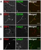
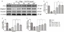
          Alzheimer’s disease (AD) is the most common form of senile dementia which is characterized by abnormal amyloid beta (Aβ) accumulation and deposition in brain parenchyma and cerebral capillaries, and leads to blood-brain barrier (BBB) disruption. Despite great progress in understanding the etiology of AD, the underlying pathogenic mechanism of BBB damage is still unclear, and no effective treatment has been devised. The standard Ginkgo biloba extract EGb761 has been widely used as a potential cognitive enhancer for the treatment of AD. However, the cellular mechanism underlying the effect remain to be clarified. In this study, we employed an immortalized endothelial cell line (bEnd.3) and incubation of Aβ 1–42 oligomer, to mimic a monolayer BBB model under conditions found in the AD brain. We investigated the effect of EGb761 on BBB and found that Aβ 1–42 oligomer-induced cell injury, apoptosis, and generation of intracellular reactive oxygen species (ROS), were attenuated by treatment with EGb761. Moreover, treatment of the cells with EGb761 decreased BBB permeability and increased tight junction scaffold protein levels including ZO-1, Claudin-5 and Occludin. We also found that the Aβ 1–42 oligomer-induced upregulation of the receptor for advanced glycation end-products (RAGE), which mediates Aβ cytotoxicity and plays an essential role in AD progression, was significantly decreased by treatment with EGb761. To our knowledge, we provide the first direct in vitro evidence of an effect of EGb761 on the brain endothelium exposed to Aβ 1–42 oligomer, and on the expression of tight junction (TJ) scaffold proteins and RAGE. Our results provide a new insight into a possible mechanism of action of EGb761. This study provides a rational basis for the therapeutic application of EGb761 in the treatment of AD.

          Related collections

          Most cited references34

          • Record: found
          • Abstract: found
          • Article: not found

          RAGE mediates amyloid-beta peptide transport across the blood-brain barrier and accumulation in brain.

          Amyloid-beta peptide (Abeta) interacts with the vasculature to influence Abeta levels in the brain and cerebral blood flow, providing a means of amplifying the Abeta-induced cellular stress underlying neuronal dysfunction and dementia. Systemic Abeta infusion and studies in genetically manipulated mice show that Abeta interaction with receptor for advanced glycation end products (RAGE)-bearing cells in the vessel wall results in transport of Abeta across the blood-brain barrier (BBB) and expression of proinflammatory cytokines and endothelin-1 (ET-1), the latter mediating Abeta-induced vasoconstriction. Inhibition of RAGE-ligand interaction suppresses accumulation of Abeta in brain parenchyma in a mouse transgenic model. These findings suggest that vascular RAGE is a target for inhibiting pathogenic consequences of Abeta-vascular interactions, including development of cerebral amyloidosis.
            Bookmark
            • Record: found
            • Abstract: found
            • Article: not found

            Amyloid Beta induces oxidative stress-mediated blood-brain barrier changes in capillary amyloid angiopathy.

            Cerebral amyloid angiopathy (CAA) is frequently observed in Alzheimer's disease (AD) and is characterized by deposition of amyloid beta (Aβ) in leptomeningeal and cortical brain vasculature. In 40% of AD cases, Aβ mainly accumulates in cortical capillaries, a phenomenon referred to as capillary CAA (capCAA). The aim of this study was to investigate blood-brain barrier (BBB) alterations in CAA-affected capillaries with the emphasis on tight junction (TJ) changes. First, capCAA brain tissue was analyzed for the distribution of TJs. Here, we show for the first time a dramatic loss of occludin, claudin-5, and ZO-1 in Aβ-laden capillaries surrounded by NADPH oxidase-2 (NOX-2)-positive activated microglia. Importantly, we observed abundant vascular expression of the Aβ transporter receptor for advanced glycation endproducts (RAGE). To unravel the underlying mechanism, a human brain endothelial cell line was stimulated with Aβ1-42 to analyze the effects of Aβ. We observed a dose-dependent cytotoxicity and increased ROS generation, which interestingly was reversed by administration of exogenous antioxidants, NOX-2 inhibitors, and by blocking RAGE. Taken together, our data evidently show that Aβ is toxic to brain endothelial cells via binding to RAGE and induction of ROS production, which ultimately leads to disruption of TJs and loss of BBB integrity.
              Bookmark
              • Record: found
              • Abstract: found
              • Article: found
              Is Open Access

              Amyloid Triggers Extensive Cerebral Angiogenesis Causing Blood Brain Barrier Permeability and Hypervascularity in Alzheimer's Disease

              Evidence of reduced blood-brain barrier (BBB) integrity preceding other Alzheimer's disease (AD) pathology provides a strong link between cerebrovascular angiopathy and AD. However, the “Vascular hypothesis”, holds that BBB leakiness in AD is likely due to hypoxia and neuroinflammation leading to vascular deterioration and apoptosis. We propose an alternative hypothesis: amyloidogenesis promotes extensive neoangiogenesis leading to increased vascular permeability and subsequent hypervascularization in AD. Cerebrovascular integrity was characterized in Tg2576 AD model mice that overexpress the human amyloid precursor protein (APP) containing the double missense mutations, APPsw, found in a Swedish family, that causes early-onset AD. The expression of tight junction (TJ) proteins, occludin and ZO-1, were examined in conjunction with markers of apoptosis and angiogenesis. In aged Tg2576 AD mice, a significant increase in the incidence of disrupted TJs, compared to age matched wild-type littermates and young mice of both genotypes, was directly linked to an increased microvascular density but not apoptosis, which strongly supports amyloidogenic triggered hypervascularity as the basis for BBB disruption. Hypervascularity in human patients was corroborated in a comparison of postmortem brain tissues from AD and controls. Our results demonstrate that amylodogenesis mediates BBB disruption and leakiness through promoting neoangiogenesis and hypervascularity, resulting in the redistribution of TJs that maintain the barrier and thus, provides a new paradigm for integrating vascular remodeling with the pathophysiology observed in AD. Thus the extensive angiogenesis identified in AD brain, exhibits parallels to the neovascularity evident in the pathophysiology of other diseases such as age-related macular degeneration.
                Bookmark

                Author and article information

                Contributors
                Role: Editor
                Journal
                PLoS One
                PLoS ONE
                plos
                plosone
                PLoS ONE
                Public Library of Science (San Francisco, USA )
                1932-6203
                2014
                26 November 2014
                : 9
                : 11
                : e113126
                Affiliations
                [1 ]Department of Neurology, Zhongshan Hospital, Fudan University, Shanghai, China
                [2 ]State Key Laboratory of Medical Neurobiology, School of Basic Medical Sciences, Fudan University, Shanghai, China
                [3 ]Geriatrics Department of Traditional Chinese Medicine, Huadong Hospital, Fudan University, Shanghai, China
                [4 ]Department of Perinatal Medicine Pregnancy Research Centre and University of Melbourne Department of Obstetrics and Gynaecology, Royal Women’s Hospital, Parkville, VIC, Australia
                [5 ]Shanghai Geriatric Institute of Chinese Medicine, Shanghai, China
                [6 ]School of Acupuncture, Massage and Rehabilitation, Yunnan University of Traditional Chinese Medicine, Kunming, China
                [7 ]Shanghai Institute of Geriatrics, Huadong Hospital, Fudan University, Shanghai, China
                University of Maryland School of Pharmacy, United States of America
                Author notes

                Competing Interests: The authors have declared that no competing interests exist.

                Conceived and designed the experiments: WBW YML SJX. Performed the experiments: WBW. Analyzed the data: LC BK CC XTT. Contributed reagents/materials/analysis tools: LC LML. Contributed to the writing of the manuscript: WBW.

                Article
                PONE-D-14-23312
                10.1371/journal.pone.0113126
                4245095
                25426944
                b3558f85-ac5f-4afa-9808-2205014cebfb
                Copyright @ 2014

                This is an open-access article distributed under the terms of the Creative Commons Attribution License, which permits unrestricted use, distribution, and reproduction in any medium, provided the original author and source are credited.

                History
                : 25 May 2014
                : 20 October 2014
                Page count
                Pages: 8
                Funding
                The authors gratefully acknowledge the financial support of this study by National Natural Science Foundation of China (Grant No. 81473739), and Shanghai Committee of Science and Technology, China (Grant No. 12401904500), YML received the funding, ( http://www.nsfc.gov.cn/) and ( http://www.stcsm.gov.cn/), respectively; by National Natural Science Foundation of China (Grant No. 31171129, Grant No. 81460748), SJX received the funding, ( http://www.nsfc.gov.cn/). The funders had no role in study design, data collection and analysis, decision to publish, or preparation of the manuscript.
                Categories
                Research Article
                Medicine and Health Sciences
                Mental Health and Psychiatry
                Dementia
                Alzheimer Disease
                Neurology
                Neurodegenerative Diseases
                Neuropharmacology
                Custom metadata
                The authors confirm that all data underlying the findings are fully available without restriction. All relevant data are within the paper.

                Uncategorized
                Uncategorized

                Comments

                Comment on this article