16
views
0
recommends
+1 Recommend
0 collections
    0
    shares
      • Record: found
      • Abstract: found
      • Article: found
      Is Open Access

      F-actin-rich contractile endothelial pores prevent vascular leakage during leukocyte diapedesis through local RhoA signalling

      research-article

      Read this article at

      Bookmark
          There is no author summary for this article yet. Authors can add summaries to their articles on ScienceOpen to make them more accessible to a non-specialist audience.

          Abstract

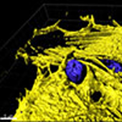
          During immune surveillance and inflammation, leukocytes exit the vasculature through transient openings in the endothelium without causing plasma leakage. However, the exact mechanisms behind this intriguing phenomenon are still unknown. Here we report that maintenance of endothelial barrier integrity during leukocyte diapedesis requires local endothelial RhoA cycling. Endothelial RhoA depletion in vitro or Rho inhibition in vivo provokes neutrophil-induced vascular leakage that manifests during the physical movement of neutrophils through the endothelial layer. Local RhoA activation initiates the formation of contractile F-actin structures that surround emigrating neutrophils. These structures that surround neutrophil-induced endothelial pores prevent plasma leakage through actomyosin-based pore confinement. Mechanistically, we found that the initiation of RhoA activity involves ICAM-1 and the Rho GEFs Ect2 and LARG. In addition, regulation of actomyosin-based endothelial pore confinement involves ROCK2b, but not ROCK1. Thus, endothelial cells assemble RhoA-controlled contractile F-actin structures around endothelial pores that prevent vascular leakage during leukocyte extravasation.

          Abstract

          Endothelial cells can support leukocyte extravasation without causing vascular leakage, but the exact mechanism underlying this process has not been fully elucidated. Here the authors show that it is regulated through actomyosin-based endothelial pore confinement, which requires local endothelial RhoA activation.

          Related collections

          Most cited references50

          • Record: found
          • Abstract: found
          • Article: not found

          Spatiotemporal dynamics of RhoA activity in migrating cells.

          Rho family GTPases regulate the actin and adhesion dynamics that control cell migration. Current models postulate that Rac promotes membrane protrusion at the leading edge and that RhoA regulates contractility in the cell body. However, there is evidence that RhoA also regulates membrane protrusion. Here we use a fluorescent biosensor, based on a novel design preserving reversible membrane interactions, to visualize the spatiotemporal dynamics of RhoA activity during cell migration. In randomly migrating cells, RhoA activity is concentrated in a sharp band directly at the edge of protrusions. It is observed sporadically in retracting tails, and is low in the cell body. RhoA activity is also associated with peripheral ruffles and pinocytic vesicles, but not with dorsal ruffles induced by platelet-derived growth factor (PDGF). In contrast to randomly migrating cells, PDGF-induced membrane protrusions have low RhoA activity, potentially because PDGF strongly activates Rac, which has previously been shown to antagonize RhoA activity. Our data therefore show that different extracellular cues induce distinct patterns of RhoA signalling during membrane protrusion.
            Bookmark
            • Record: found
            • Abstract: found
            • Article: not found

            Leukocyte extravasation and vascular permeability are each controlled in vivo by different tyrosine residues of VE-cadherin.

            Tyrosine phosphorylation of the adhesion molecule VE-cadherin is assumed to affect endothelial junction integrity. However, it remains unclear whether tyrosine residues of VE-cadherin are required for the induction of vascular permeability and the regulation of leukocyte extravasation in vivo. We found here that knock-in mice expressing a Y685F mutant of VE-cadherin had impaired induction of vascular permeability, but those expressing a Y731F mutant did not. In contrast, mice expressing the Y731F VE-cadherin mutant showed decreased neutrophil-extravasation in cremaster tissue, but those expressing the Y685F mutant did not. Whereas inflammatory mediators induced the phosphorylation of Tyr685 in vivo, Tyr731 showed high baseline phosphorylation. Leukocytes triggered dephosphorylation of Tyr731 via the tyrosine phosphatase SHP-2, which allowed the adaptin AP-2 to bind and initiate endocytosis of VE-cadherin. Thus, Tyr685 and Tyr731 of VE-cadherin distinctly and selectively regulate the induction of vascular permeability or leukocyte extravasation.
              Bookmark
              • Record: found
              • Abstract: not found
              • Article: not found

              Lifeact mice for studying F-actin dynamics.

                Bookmark

                Author and article information

                Journal
                Nat Commun
                Nat Commun
                Nature Communications
                Nature Publishing Group
                2041-1723
                27 January 2016
                2016
                : 7
                : 10493
                Affiliations
                [1 ]Department of Molecular Cell Biology, Sanquin Research and Landsteiner Laboratory, Academic Medical Centre, University of Amsterdam , 1066CX Amsterdam, The Netherlands
                [2 ]Genetics and Developmental Biology, Center for Cell Analyses and Modelling, University of Connecticut Health Centre , Farmington, Connecticut 06032, USA
                [3 ]Experimental Medicine and Pharmacology, Centre for Microvascular Research, William Harvey Research Institute, Barts and The London School of Medicine and Dentistry, Queen Mary, University of London , Charterhouse Square, London, EC1M 6BQ, UK
                [4 ]Molecular Cytology, Swammerdam Institute for Life Sciences, University of Amsterdam , Amsterdam 1098XH, The Netherlands
                [5 ]Department of Angiogenesis, Walter-Brendel-Center of Experimental Medicine Ludwig-Maximilians University Marchioninistr. 27 81377 Munich, Germany
                Author notes
                Author information
                http://orcid.org/0000-0002-1091-475X
                http://orcid.org/0000-0002-0630-3825
                Article
                ncomms10493
                10.1038/ncomms10493
                4737874
                26814335
                b3a47fe6-f2b6-4234-afbc-02f3601f40af
                Copyright © 2016, Nature Publishing Group, a division of Macmillan Publishers Limited. All Rights Reserved.

                This work is licensed under a Creative Commons Attribution 4.0 International License. The images or other third party material in this article are included in the article's Creative Commons license, unless indicated otherwise in the credit line; if the material is not included under the Creative Commons license, users will need to obtain permission from the license holder to reproduce the material. To view a copy of this license, visit http://creativecommons.org/licenses/by/4.0/

                History
                : 28 April 2015
                : 14 December 2015
                Categories
                Article

                Uncategorized
                Uncategorized

                Comments

                Comment on this article