10
views
0
recommends
+1 Recommend
0 collections
    0
    shares
      • Record: found
      • Abstract: found
      • Article: found
      Is Open Access

      Meta-analysis of the clinical and immunopathological characteristics and treatment outcomes in epidermolysis bullosa acquisita patients

      research-article

      Read this article at

      Bookmark
          There is no author summary for this article yet. Authors can add summaries to their articles on ScienceOpen to make them more accessible to a non-specialist audience.

          Abstract

          Background

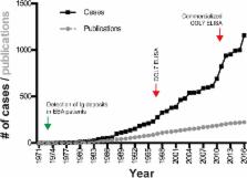
          Epidermolysis bullosa acquisita (EBA) is an orphan autoimmune disease. Several clinical phenotypes have been described, but subepidermal blistering is characteristic of all variants. Limited data on clinical and immunopathological characteristics and treatment outcomes in EBA are available. To fill this gap, we collected this information from EBA cases, meeting current diagnostic criteria, published between 1971 and 2016.

          Results

          We identified 1159 EBA cases. This number must be, however, interpreted with caution, as it is not possible to check for multiple reporting. The analysis of all cases indicated that EBA affects all age groups (median: 50 years, range: 1 to 94 years) at an equal gender distribution. Non-mechanobullous (non-MB) forms of EBA were observed in 55% of patients, whereas the mechanobullous variant (MB-EBA) or a combination of both variants was described in 38 or 7% of patients, respectively. Type VII collagen (COL7)-specific autoantibodies were primarily of the IgG isotype, but anti-COL7 IgA, IgM and IgE were also documented. Comparison of the 2 clinical EBA types showed a higher frequency of IgA deposits in non-MB EBA as opposed to MB EBA. Mucous membrane involvement was observed in 23% of patients, and 4.4% of cases were associated with other chronic inflammatory diseases. Of note, IgA deposits were more frequently observed in cases with mucous membrane involvement. Our analysis indicated that EBA is difficult to treat and that the choice of treatment varies widely. Chi square was applied to identify medications associated with complete remission (CR). Considering all EBA cases, intravenous immunoglobulin (IVIG, p = 0.0047) and rituximab ( p = 0.0114) were associated with CR. Subgroup analysis demonstrated that no treatment was associated with CR for non-MB EBA, while IVIG ( p = 0.003) was associated with CR in MB EBA.

          Conclusions

          Within the limitations of the study , we here document the clinical and immunopathological characteristics and treatment outcomes in a large cohort of EBA patients. The observed associations of single drugs with treatment outcome may serve as a guide to develop clinical trials.

          Electronic supplementary material

          The online version of this article (10.1186/s13023-018-0896-1) contains supplementary material, which is available to authorized users.

          Related collections

          Most cited references66

          • Record: found
          • Abstract: found
          • Article: not found

          Epidemiology of autoimmune diseases in Denmark.

          An epidemiologic study of the autoimmune diseases taken together has not been done heretofore. The National Patient Register of Denmark is used to estimate the population prevalence of 31 possible or probable autoimmune diseases. Record linkage is used to estimate 465 pairwise co-morbidities in individuals among the 31 diseases, and familial aggregation among sibs, parents and offspring. The prevalence of any of the 31 diseases in the population is more than 5%. Within individuals, there is extensive comorbidity across the 31 diseases. Within families, aggregation is strongest for individual diseases and weak across diseases. These data confirm the importance of the autoimmune diseases as a group and suggest that common etiopathologies exist among them.
            Bookmark
            • Record: found
            • Abstract: not found
            • Article: not found

            Range of antinuclear antibodies in “healthy” individuals

              Bookmark
              • Record: found
              • Abstract: found
              • Article: not found

              Identification of the skin basement-membrane autoantigen in epidermolysis bullosa acquisita.

              Epidermolysis bullosa acquisita is an acquired chronic blistering disease of the skin, in which separation of the skin occurs in the basement-membrane zone between the epidermis and the dermis. There is evidence that blistering is initiated by an immune process. Using serum samples from nine patients as a source of antibodies, we have identified a major protein of the basement membrane of human skin that serves as the antigen (or target) for autoantibodies in this disorder. This previously unrecognized protein, which consists of two components of 290,000 and 145,000 daltons, is distinct from other known components of the basement membrane. These studies provide evidence that epidermolysis bullosa acquisita is a specific disease that is different from other primary bullous diseases, such as bullous pemphigoid and pemphigus vulgaris, and suggest that the basement-membrane component that has been identified may have a role in normal epidermal-dermal adherence.
                Bookmark

                Author and article information

                Contributors
                hiroaki.iwata@med.hokudai.ac.jp
                artem.vorobyev@uksh.de
                hiroshi_koga@med.kurume-u.ac.jp
                andreas.recke@uksh.de
                detlef.zillikens@uksh.de
                catherine.prost@aphp.fr
                norito@med.kurume-u.ac.jp
                hashyt@gmail.com
                +49 451 500-41686 , ralf.ludwig@uksh.de
                Journal
                Orphanet J Rare Dis
                Orphanet J Rare Dis
                Orphanet Journal of Rare Diseases
                BioMed Central (London )
                1750-1172
                4 September 2018
                4 September 2018
                2018
                : 13
                : 153
                Affiliations
                [1 ]ISNI 0000 0001 0057 2672, GRID grid.4562.5, Department of Dermatology, , University of Lübeck, ; Ratzeburger Allee 160, D-23538 Lübeck, Germany
                [2 ]ISNI 0000 0001 0706 0776, GRID grid.410781.b, Department of Dermatology, , Kurume University School of Medicine, and Kurume University Institute of Cutaneous Cell Biology, ; Kurume, Fukuoka, Japan
                [3 ]ISNI 0000 0000 8715 2621, GRID grid.413780.9, Referral center for auto-immune bullous diseases, Department of Dermatology, APHP, Avicenne Hospital, ; Bobigny, France
                [4 ]ISNI 0000 0001 1009 6411, GRID grid.261445.0, Department of Dermatology, Faculty of Medicine, , Osaka City University, ; Osaka, Japan
                [5 ]ISNI 0000 0001 2173 7691, GRID grid.39158.36, Present address: Department of Dermatology, , Hokkaido University Graduate School of Medicine, ; Sapporo, Japan
                [6 ]ISNI 0000 0001 0057 2672, GRID grid.4562.5, Lübeck Institute of Experimental Dermatology, University of Lübeck, ; Lübeck, Germany
                Author information
                http://orcid.org/0000-0002-1394-1737
                Article
                896
                10.1186/s13023-018-0896-1
                6122731
                30180870
                b3ea4ce7-eaa4-4c3b-9f4b-dc0629fe072c
                © The Author(s). 2018

                Open Access This article is distributed under the terms of the Creative Commons Attribution 4.0 International License ( http://creativecommons.org/licenses/by/4.0/), which permits unrestricted use, distribution, and reproduction in any medium, provided you give appropriate credit to the original author(s) and the source, provide a link to the Creative Commons license, and indicate if changes were made. The Creative Commons Public Domain Dedication waiver ( http://creativecommons.org/publicdomain/zero/1.0/) applies to the data made available in this article, unless otherwise stated.

                History
                : 8 June 2017
                : 22 August 2018
                Funding
                Funded by: FundRef http://dx.doi.org/10.13039/501100001659, Deutsche Forschungsgemeinschaft;
                Award ID: EXC 306/2
                Award ID: GRK 1727/2
                Award ID: GRK 1743/2
                Award ID: KFO 303/1
                Award Recipient :
                Categories
                Research
                Custom metadata
                © The Author(s) 2018

                Infectious disease & Microbiology
                epidermolysis bullosa acquisita,treatment,meta-analysis,diagnosis,ivig,rituximab

                Comments

                Comment on this article