4
views
0
recommends
+1 Recommend
0 collections
    0
    shares
      • Record: found
      • Abstract: found
      • Article: not found

      Regulation of Lipolytic Response and Energy Balance by Melanocortin 2 Receptor Accessory Protein (MRAP) in Adipocytes

      research-article

      Read this article at

      ScienceOpenPublisherPMC
      Bookmark
          There is no author summary for this article yet. Authors can add summaries to their articles on ScienceOpen to make them more accessible to a non-specialist audience.

          Abstract

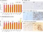
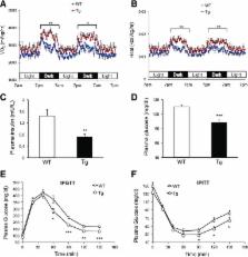
          Melanocortin 2 receptor accessory protein (MRAP) is highly expressed in adrenal gland and adipose tissue. In adrenal cells, MRAP is essential for adrenocorticotropic hormone (ACTH)–induced activation of the cAMP/protein kinase A (PKA) pathway by melanocortin 2 receptor (MC2R), leading to glucocorticoid production and secretion. Although ACTH was known to stimulate PKA-dependent lipolysis, the functional involvement of MRAP in adipocyte metabolism remains incompletely defined. Herein, we found that knockdown or overexpression of MRAP in 3T3-L1 adipocytes reduced or increased ACTH-induced lipolysis, respectively. Moreover, an unbiased proteomics screen and coimmunoprecipitation analysis identified Gαs as a novel interacting partner of MRAP. An MRAP mutant disabled in Gαs association failed to augment the activation of PKA and lipolytic response to ACTH. Furthermore, compared with wild-type mice, transgenic mice (aP2-MRAP) overexpressing MRAP fat specifically exhibited increased lipolytic response to ACTH. When fed a high-fat diet (HFD), the transgenic mice displayed a significant decrease in the gain of adiposity and body weight as well as an improvement in glucose and insulin tolerance. These phenotypes were accompanied by increased adipose expression of genes for mitochondrial fatty acid oxidation and thermogenesis, and overall energy expenditure. Collectively, our data strongly suggest that MRAP plays a critical role in the regulation of ACTH-induced adipose lipolysis and whole-body energy balance.

          Related collections

          Most cited references33

          • Record: found
          • Abstract: found
          • Article: not found

          Fat mobilization in adipose tissue is promoted by adipose triglyceride lipase.

          Mobilization of fatty acids from triglyceride stores in adipose tissue requires lipolytic enzymes. Dysfunctional lipolysis affects energy homeostasis and may contribute to the pathogenesis of obesity and insulin resistance. Until now, hormone-sensitive lipase (HSL) was the only enzyme known to hydrolyze triglycerides in mammalian adipose tissue. Here, we report that a second enzyme, adipose triglyceride lipase (ATGL), catalyzes the initial step in triglyceride hydrolysis. It is interesting that ATGL contains a "patatin domain" common to plant acyl-hydrolases. ATGL is highly expressed in adipose tissue of mice and humans. It exhibits high substrate specificity for triacylglycerol and is associated with lipid droplets. Inhibition of ATGL markedly decreases total adipose acyl-hydrolase activity. Thus, ATGL and HSL coordinately catabolize stored triglycerides in adipose tissue of mammals.
            Bookmark
            • Record: found
            • Abstract: found
            • Article: not found

            FAT SIGNALS - Lipases and Lipolysis in Lipid Metabolism and Signaling

            Lipolysis is defined as the catabolism of triacylglycerols stored in cellular lipid droplets. Recent discoveries of essential lipolytic enzymes and characterization of numerous regulatory proteins and mechanisms have fundamentally changed our perception of lipolysis and its impact on cellular metabolism. New findings that lipolytic products and intermediates participate in cellular signaling processes and that “lipolytic signaling” is particularly important in many nonadipose tissues unveil a previously underappreciated aspect of lipolysis, which may be relevant for human disease.
              Bookmark
              • Record: found
              • Abstract: found
              • Article: found
              Is Open Access

              Glucocorticoids Acutely Increase Brown Adipose Tissue Activity in Humans, Revealing Species-Specific Differences in UCP-1 Regulation

              Summary The discovery of brown adipose tissue (BAT) in adult humans presents a new therapeutic target for metabolic disease; however, little is known about the regulation of human BAT. Chronic glucocorticoid excess causes obesity in humans, and glucocorticoids suppress BAT activation in rodents. We tested whether glucocorticoids regulate BAT activity in humans. In vivo, the glucocorticoid prednisolone acutely increased 18fluorodeoxyglucose uptake by BAT (measured using PET/CT) in lean healthy men during mild cold exposure (16°C–17°C). In addition, prednisolone increased supraclavicular skin temperature (measured using infrared thermography) and energy expenditure during cold, but not warm, exposure in lean subjects. In vitro, glucocorticoids increased isoprenaline-stimulated respiration and UCP-1 in human primary brown adipocytes, but substantially decreased isoprenaline-stimulated respiration and UCP-1 in primary murine brown and beige adipocytes. The highly species-specific regulation of BAT function by glucocorticoids may have important implications for the translation of novel treatments to activate BAT to improve metabolic health.
                Bookmark

                Author and article information

                Journal
                Diabetes
                Diabetes
                diabetes
                diabetes
                Diabetes
                Diabetes
                American Diabetes Association
                0012-1797
                1939-327X
                February 2018
                07 December 2017
                : 67
                : 2
                : 222-234
                Affiliations
                [1] 1Department of Biochemistry and Molecular Biology, Mayo Clinic College of Medicine, Scottsdale, AZ
                [2] 2HEALth Program, Mayo Clinic, Scottsdale, AZ
                [3] 3School of Life Sciences, Arizona State University, Tempe, AZ
                [4] 4Division of Endocrinology, Mayo Clinic, Scottsdale, AZ
                Author notes
                Corresponding author: Jun Liu, liu.jun@ 123456mayo.edu .
                Author information
                http://orcid.org/0000-0002-3646-0004
                Article
                0862
                10.2337/db17-0862
                5780064
                29217655
                b5b8ee41-bdd9-4c6d-93a3-642946d04696
                © 2017 by the American Diabetes Association.

                Readers may use this article as long as the work is properly cited, the use is educational and not for profit, and the work is not altered. More information is available at http://www.diabetesjournals.org/content/license.

                History
                : 26 July 2017
                : 22 November 2017
                Page count
                Figures: 7, Tables: 0, Equations: 0, References: 39, Pages: 13
                Funding
                Funded by: Arizona Biomedical Research Commission, DOI http://dx.doi.org/10.13039/100008335;
                Award ID: ADHS14-00003606
                Funded by: National Institute of Diabetes and Digestive and Kidney Diseases, DOI http://dx.doi.org/10.13039/100000062;
                Award ID: DK089178
                Award ID: DK109096
                Categories
                0601
                Metabolism

                Endocrinology & Diabetes
                Endocrinology & Diabetes

                Comments

                Comment on this article