250
views
0
recommends
+1 Recommend
1 collections
    0
    shares
      • Record: found
      • Abstract: found
      • Article: found
      Is Open Access

      Prospective evaluation of the proportion of sessile serrated adenoma/polyps in endoscopically diagnosed colorectal polyps with hyperplastic features1

      research-article

      Read this article at

      ScienceOpenPublisherPMC
      Bookmark
          There is no author summary for this article yet. Authors can add summaries to their articles on ScienceOpen to make them more accessible to a non-specialist audience.

          Abstract

          Background and study aims: Sessile serrated adenoma/polyps (SSA/Ps) are considered precursors of colorectal cancers with microsatellite instability. However, it is still difficult to differentiate SSA/Ps from hyperplastic polyps endoscopically; therefore, the prevalence of SSA/Ps remains uncertain in clinical practice. This study aimed to clarify the proportion of SSA/Ps in endoscopically diagnosed colorectal polyps with hyperplastic features (E-HPs).

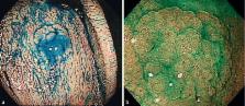
          Patients and methods: Patients aged ≥ 40 years undergoing colonoscopy for standard clinical indications at our center were prospectively enrolled between June 2013 and May 2014. During colonoscopy, 0.05 % indigo carmine dye was sprayed throughout the colorectum to highlight lesions. All detected lesions were diagnosed by high definition magnifying narrow-band imaging and were resected endoscopically or surgically, apart from rectosigmoid E-HPs ≤ 5 mm. The number of rectosigmoid E-HPs ≤ 5 mm was recorded, and some were resected for use as tissue samples.

          Results: A total of 343 patients (male: 42.9 %; mean age: 61.5 years) were included. Among 3838 E-HPs (distal: 96.4 %) detected in 294 patients, 792 were resected and analyzed. All of 21 SSA/Ps identified in 17 patients were included in E-HPs, and the overall proportion of SSA/Ps in E-HPs was 2.7 %. However, this proportion increased with the size of E-HPs (≤ 5 mm: 0.7 %; 6 – 9 mm: 29.0 %; ≥ 10 mm: 70 %) and was higher in the proximal colon than in the distal colorectum (10.9 % vs. 0.9 %). In addition, no SSA/P was found in the rectum, and no SSA/P had cytological dysplasia.

          Conclusions: The overall proportion of SSA/Ps in E-HPs was 2.7 %, although this proportion was higher in the proximal colon and increased with the size of E-HPs. SSA/Ps were common in routine colonoscopy, with a prevalence of at least 5.0 %.

          Study registration: UMIN000010832.

          Most cited references24

          • Record: found
          • Abstract: found
          • Article: not found

          Serrated lesions of the colorectum: review and recommendations from an expert panel.

          Serrated lesions of the colorectum are the precursors of perhaps one-third of colorectal cancers (CRCs). Cancers arising in serrated lesions are usually in the proximal colon, and account for a disproportionate fraction of cancer identified after colonoscopy. We sought to provide guidance for the clinical management of serrated colorectal lesions based on current evidence and expert opinion regarding definitions, classification, and significance of serrated lesions. A consensus conference was held over 2 days reviewing the topic of serrated lesions from the perspectives of histology, molecular biology, epidemiology, clinical aspects, and serrated polyposis. Serrated lesions should be classified pathologically according to the World Health Organization criteria as hyperplastic polyp, sessile serrated adenoma/polyp (SSA/P) with or without cytological dysplasia, or traditional serrated adenoma (TSA). SSA/P and TSA are premalignant lesions, but SSA/P is the principal serrated precursor of CRCs. Serrated lesions have a distinct endoscopic appearance, and several lines of evidence suggest that on average they are more difficult to detect than conventional adenomatous polyps. Effective colonoscopy requires an endoscopist trained in the endoscopic appearance of serrated lesions. We recommend that all serrated lesions proximal to the sigmoid colon and all serrated lesions in the rectosigmoid > 5 mm in size, be completely removed. Recommendations are made for post-polypectomy surveillance of serrated lesions and for surveillance of serrated polyposis patients and their relatives.
            Bookmark
            • Record: found
            • Abstract: found
            • Article: not found

            Validation of a simple classification system for endoscopic diagnosis of small colorectal polyps using narrow-band imaging.

            Almost all colorectal polyps ≤ 5 mm are benign, yet current practice requires costly pathologic analysis. We aimed to develop and evaluate the validity of a simple narrow-band imaging (NBI)-based classification system for differentiating hyperplastic from adenomatous polyps. The study was conducted in 4 phases: (1) evaluation of accuracy and reliability of histologic prediction by NBI-experienced colonoscopists; (2) development of a classification based on color, vessels, and surface pattern criteria, using a modified Delphi method; (3) validation of the component criteria by people not experienced in endoscopy or NBI analysis (25 medical students, 19 gastroenterology fellows) using 118 high-definition colorectal polyp images of known histology; and (4) validation of the classification system by NBI-trained gastroenterology fellows, using still images. We performed a pilot evaluation during real-time colonoscopy. We developed a classification system for the endoscopic diagnosis of colorectal polyp histology and established its predictive validity. When all 3 criteria were used, the specificity ranged from 94.9% to 100% and the combined sensitivity ranged from 8.5% to 61.0%. The specificities of the individual criteria were lower although the sensitivities were higher. During real-time colonoscopy, endoscopists made diagnoses with high confidence for 75% of consecutive small colorectal polyps, with 89% accuracy, 98% sensitivity, and 95% negative predictive values. We developed and established the validity of an NBI classification system that can be used to diagnose colorectal polyps. In preliminary real-time evaluation, the system allowed endoscopic diagnoses of colorectal polyp histology. Copyright © 2012 AGA Institute. Published by Elsevier Inc. All rights reserved.
              Bookmark
              • Record: found
              • Abstract: found
              • Article: not found

              Endoscopic prediction of deep submucosal invasive carcinoma: validation of the narrow-band imaging international colorectal endoscopic (NICE) classification.

              A simple endoscopic classification to accurately predict deep submucosal invasive (SM-d) carcinoma would be clinically useful. To develop and assess the validity of the NBI international colorectal endoscopic (NICE) classification for the characterization of SM-d carcinoma. The study was conducted in 4 phases: (1) evaluation of endoscopic differentiation by NBI-experienced colonoscopists; (2) extension of the NICE classification to incorporate SM-d (type 3) by using a modified Delphi method; (3) prospective validation of the individual criteria by inexperienced participants, by using high-definition still images without magnification of known histology; and (4) prospective validation of the individual criteria and overall classification by inexperienced participants after training. Japanese academic unit. Performance characteristics of the NICE criteria (phase 3) and overall classification (phase 4) for SM-d carcinoma; sensitivity, specificity, predictive values, and accuracy. We expanded the NICE classification for the endoscopic diagnosis of SM-d carcinoma (type 3) and established the predictive validity of its individual components. The negative predictive values of the individual criteria for diagnosis of SM-d carcinoma were 76.2% (color), 88.5% (vessels), and 79.1% (surface pattern). When any 1 of the 3 SM-d criteria was present, the sensitivity was 94.9%, and the negative predictive value was 95.9%. The overall sensitivity and negative predictive value of a global, high-confidence prediction of SM-d carcinoma was 92%. Interobserver agreement for an overall SM-d carcinoma prediction was substantial (kappa 0.70). Single Japanese center, use of still images without prospective clinical evaluation. The NICE classification is a valid tool for predicting SM-d carcinomas in colorectal tumors. Copyright © 2013 American Society for Gastrointestinal Endoscopy. Published by Mosby, Inc. All rights reserved.
                Bookmark

                Author and article information

                Journal
                Endosc Int Open
                Endosc Int Open
                10.1055/s-0034-1377934
                Endoscopy International Open
                © Georg Thieme Verlag KG (Stuttgart · New York )
                2364-3722
                2196-9736
                August 2015
                05 May 2015
                : 3
                : 4
                : E354-E358
                Affiliations
                [1 ]Gastrointestinal Center, Sano Hospital, Hyogo, Japan
                [2 ]Department of Pathology, Shinko Hospital, Hyogo, Japan
                Author notes
                Corresponding author Wataru Sano, MD Gastrointestinal Center Sano Hospital 2-5-1 ShimizugaokaTarumiKobeHyogo 655-0031Japan+81-78-7850077 watasano@ 123456yahoo.co.jp
                Article
                10.1055/s-0034-1391948
                4554512
                26357681
                b8e0ea23-5abe-42fb-bdcc-583ab69323fd
                © Thieme Medical Publishers
                History
                : 28 November 2014
                : 02 March 2015
                Categories
                Article

                Comments

                Comment on this article