15
views
0
recommends
+1 Recommend
2 collections
    0
    shares
      • Record: found
      • Abstract: found
      • Article: not found

      Transient cortical blindness in COVID-19 pneumonia; a PRES-like syndrome: A case report

      letter

      Read this article at

      ScienceOpenPublisherPMC
      Bookmark
          There is no author summary for this article yet. Authors can add summaries to their articles on ScienceOpen to make them more accessible to a non-specialist audience.

          Abstract

          Dear Editor, The World Health Organization declared the outbreak of the 2019 novel coronavirus, in March 12th, 2020, a global pandemic after widely spreading of the epidemic COVID-19 pneumonia cases [1]. It has been reported that, in addition to the respiratory tract infection symptoms, patients can also have neurologic signs and symptoms; like acute cerebrovascular disease, polyneuritis, encephalitis and encephalopathy [2]. In this report, we describe a patient who developed bilateral reversible cortical blindness, who presented by COVID-19 related pneumonia. A 38 years old male patient, admitted to emergency department with a history of fever for 5 days. His body temperature was 38,5 °C, blood pressure was 130/80 mmHg and oxygen saturation 98% while he was breathing ambient air. Breath sounds were normal with adventitious sounds on both sides. His chest CT scan showed multiple, multilobar, peripheral ground-glass opasifications in both lungs (Fig. 1 ). Laboratory tests results showed highly elevated CRP and ferritin levels with marked lymphopenia. His nasopharyngeal swab reverse transcription-PCR (RT-PCR) was positive for SARS-CoV-2. After admission, he received hydroxychloroquine (400 mg for the first day, 200 mg/day for four days), azitromicin 500 mg/day, and osetalmivir 150 mg/day combined with nasal oxygen therapy. His oxygen saturation was declined to 88% on the second day and non-invasive mechanical ventilatory support was started at intensive care unit (ICU). On the fifth day of ICU, he suddenly developed acute confusional state with agitation and his blood pressure observed to be at high levels for a few hours. Meanwhile, the patient complained about vision loss in both eyes. In his neurological examination he was awake, but apathic and hardly obeying commands. His pupils were 2 mm and equally reactive to light. Fundus examination was normal. His visual acuity was severely impared on both eyes; he could only recognize waving hands and there was perception of light. His entire neurological examination was normal. Brain magnetic resonance imaging (MRI) showed bilateral, especially left occipital, frontal cortical white matter and splenium of corpus callosum T2/Fluid-attenuated inversion recovery (FLAIR) hyperintensities and diffusion restriction in diffusion weighted imaging (DWI) (Fig. 2 ) revealing vasogenic edema similar to posterior reversible leucoencephalopathy (PRES). Hydroxychloroquine treatment was stopped and the dexamethasone with a 24 mg/day dose is started. On second dose of corticosteroid treatment, patient was able to obey commands and his visual impairment fully recovered. In his neurocognitive assessment we determined visual agnosia which lasted in a week. The corticosteroid therapy tapered and stopped in two weeks' time. His neurological examination and neurocognitive assessment were completely normal on the tenth day. The brain MRI performed two weeks later, showed complete regression of the lesions (Fig. 3 ). Fig. 1 Torax CT showed multiple, dominantly right patchy, peripheral, ground-glass opasities in both lungs a) coronal, b) Axial images. Fig. 1 Fig. 2 Brain diffusion weighted (DWI) MRI (a) showed bilateral, especially left occipital, frontal cortical white matter and splenium of corpus callosum diffusion restriction, (b) Apparent Diffusion Coefficient (ADC) showed reduced ADC values due to vasogenic edema and FLAIR sequences (c) showed hiperinteinsities in the same localizations. Fig. 2 Fig. 3 On the second week of therapy DWI (a), ADC (b) and FLAIR (c) sequences showed complete regression of the lesions. Fig. 3 1 Discussion We still don't know why focal neurological deficits may arise during SARS-CoV-2 infection. Common suggestions for pathological mechanisms are direct virus infection invasion or inflamatory factors. Recent autopsy reports have revealed that, like many viral infections SARS-CoV-2 can cause brain tissue edema and partial neuronal degeneration 
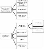
[3]. Infectious toxic encephalopathy is a reversible brain dysfunction syndrome caused by systemic toxemia, metabolic disorders and hypoxia during the process of acute infection [4]. In this disease, main pathological change is brain edema without evidence of inflammation on cerebrospinal fluid analysis. Hypoxia in the brain causes anaerobic metabolism in the mitochondria of neurons and this leads cerebral vasodilatation, swelling of neurons, interstitial edema and obstruction of cerebral blood flow [5]. PRES is a result of a systemic inflammatory state causing endothelial dysfunction [6]. That hypothesis is supported by the observation that PRES is usually associated with a systemic inflammatory process such as sepsis, eclampsia, transplantation, and autoimmune disease [7]. Although we could not determine the exact ethiology in our case, regulating the blood pressure controlling the vasogenic edema by corticosteroid treatment and controlling the virus related pneumonia have helped fort he recovery of our patient. Unfortunately, evidences are lacking to determine which of these features were due to infectious toxic encephalopathy, and which features were specific to SARS-CoV-2 infection. Declaration of Competing Interest The authors declare that there are no competing interests associated with the manuscript.

          Related collections

          Most cited references6

          • Record: found
          • Abstract: found
          • Article: not found

          Pathological findings of COVID-19 associated with acute respiratory distress syndrome

          Since late December, 2019, an outbreak of a novel coronavirus disease (COVID-19; previously known as 2019-nCoV)1, 2 was reported in Wuhan, China, 2 which has subsequently affected 26 countries worldwide. In general, COVID-19 is an acute resolved disease but it can also be deadly, with a 2% case fatality rate. Severe disease onset might result in death due to massive alveolar damage and progressive respiratory failure.2, 3 As of Feb 15, about 66 580 cases have been confirmed and over 1524 deaths. However, no pathology has been reported due to barely accessible autopsy or biopsy.2, 3 Here, we investigated the pathological characteristics of a patient who died from severe infection with severe acute respiratory syndrome coronavirus 2 (SARS-CoV-2) by postmortem biopsies. This study is in accordance with regulations issued by the National Health Commission of China and the Helsinki Declaration. Our findings will facilitate understanding of the pathogenesis of COVID-19 and improve clinical strategies against the disease. A 50-year-old man was admitted to a fever clinic on Jan 21, 2020, with symptoms of fever, chills, cough, fatigue and shortness of breath. He reported a travel history to Wuhan Jan 8–12, and that he had initial symptoms of mild chills and dry cough on Jan 14 (day 1 of illness) but did not see a doctor and kept working until Jan 21 (figure 1 ). Chest x-ray showed multiple patchy shadows in both lungs (appendix p 2), and a throat swab sample was taken. On Jan 22 (day 9 of illness), the Beijing Centers for Disease Control (CDC) confirmed by reverse real-time PCR assay that the patient had COVID-19. Figure 1 Timeline of disease course according to days from initial presentation of illness and days from hospital admission, from Jan 8–27, 2020 SARS-CoV-2=severe acute respiratory syndrome coronavirus 2. He was immediately admitted to the isolation ward and received supplemental oxygen through a face mask. He was given interferon alfa-2b (5 million units twice daily, atomisation inhalation) and lopinavir plus ritonavir (500 mg twice daily, orally) as antiviral therapy, and moxifloxacin (0·4 g once daily, intravenously) to prevent secondary infection. Given the serious shortness of breath and hypoxaemia, methylprednisolone (80 mg twice daily, intravenously) was administered to attenuate lung inflammation. Laboratory tests results are listed in the appendix (p 4). After receiving medication, his body temperature reduced from 39·0 to 36·4 °C. However, his cough, dyspnoea, and fatigue did not improve. On day 12 of illness, after initial presentation, chest x-ray showed progressive infiltrate and diffuse gridding shadow in both lungs. He refused ventilator support in the intensive care unit repeatedly because he suffered from claustrophobia; therefore, he received high-flow nasal cannula (HFNC) oxygen therapy (60% concentration, flow rate 40 L/min). On day 13 of illness, the patient's symptoms had still not improved, but oxygen saturation remained above 95%. In the afternoon of day 14 of illness, his hypoxaemia and shortness of breath worsened. Despite receiving HFNC oxygen therapy (100% concentration, flow rate 40 L/min), oxygen saturation values decreased to 60%, and the patient had sudden cardiac arrest. He was immediately given invasive ventilation, chest compression, and adrenaline injection. Unfortunately, the rescue was not successful, and he died at 18:31 (Beijing time). Biopsy samples were taken from lung, liver, and heart tissue of the patient. Histological examination showed bilateral diffuse alveolar damage with cellular fibromyxoid exudates (figure 2A, B ). The right lung showed evident desquamation of pneumocytes and hyaline membrane formation, indicating acute respiratory distress syndrome (ARDS; figure 2A). The left lung tissue displayed pulmonary oedema with hyaline membrane formation, suggestive of early-phase ARDS (figure 2B). Interstitial mononuclear inflammatory infiltrates, dominated by lymphocytes, were seen in both lungs. Multinucleated syncytial cells with atypical enlarged pneumocytes characterised by large nuclei, amphophilic granular cytoplasm, and prominent nucleoli were identified in the intra-alveolar spaces, showing viral cytopathic-like changes. No obvious intranuclear or intracytoplasmic viral inclusions were identified. Figure 2 Pathological manifestations of right (A) and left (B) lung tissue, liver tissue (C), and heart tissue (D) in a patient with severe pneumonia caused by SARS-CoV-2 SARS-CoV-2=severe acute respiratory syndrome coronavirus 2. The pathological features of COVID-19 greatly resemble those seen in SARS and Middle Eastern respiratory syndrome (MERS) coronavirus infection.4, 5 In addition, the liver biopsy specimens of the patient with COVID-19 showed moderate microvesicular steatosis and mild lobular and portal activity (figure 2C), indicating the injury could have been caused by either SARS-CoV-2 infection or drug-induced liver injury. There were a few interstitial mononuclear inflammatory infiltrates, but no other substantial damage in the heart tissue (figure 2D). Peripheral blood was prepared for flow cytometric analysis. We found that the counts of peripheral CD4 and CD8 T cells were substantially reduced, while their status was hyperactivated, as evidenced by the high proportions of HLA-DR (CD4 3·47%) and CD38 (CD8 39·4%) double-positive fractions (appendix p 3). Moreover, there was an increased concentration of highly proinflammatory CCR6+ Th17 in CD4 T cells (appendix p 3). Additionally, CD8 T cells were found to harbour high concentrations of cytotoxic granules, in which 31·6% cells were perforin positive, 64·2% cells were granulysin positive, and 30·5% cells were granulysin and perforin double-positive (appendix p 3). Our results imply that overactivation of T cells, manifested by increase of Th17 and high cytotoxicity of CD8 T cells, accounts for, in part, the severe immune injury in this patient. X-ray images showed rapid progression of pneumonia and some differences between the left and right lung. In addition, the liver tissue showed moderate microvesicular steatosis and mild lobular activity, but there was no conclusive evidence to support SARS-CoV-2 infection or drug-induced liver injury as the cause. There were no obvious histological changes seen in heart tissue, suggesting that SARS-CoV-2 infection might not directly impair the heart. Although corticosteroid treatment is not routinely recommended to be used for SARS-CoV-2 pneumonia, 1 according to our pathological findings of pulmonary oedema and hyaline membrane formation, timely and appropriate use of corticosteroids together with ventilator support should be considered for the severe patients to prevent ARDS development. Lymphopenia is a common feature in the patients with COVID-19 and might be a critical factor associated with disease severity and mortality. 3 Our clinical and pathological findings in this severe case of COVID-19 can not only help to identify a cause of death, but also provide new insights into the pathogenesis of SARS-CoV-2-related pneumonia, which might help physicians to formulate a timely therapeutic strategy for similar severe patients and reduce mortality. This online publication has been corrected. The corrected version first appeared at thelancet.com/respiratory on February 25, 2020
            Bookmark
            • Record: found
            • Abstract: found
            • Article: not found

            Posterior reversible encephalopathy syndrome, part 2: controversies surrounding pathophysiology of vasogenic edema.

            Posterior reversible encephalopathy syndrome (PRES) is a neurotoxic state accompanied by a unique brain imaging pattern typically associated with a number of complex clinical conditions including: preeclampsia/eclampsia, allogeneic bone marrow transplantation, solid organ transplantation, autoimmune diseases and high dose cancer chemotherapy. The mechanism behind the developing vasogenic edema and CT or MR imaging appearance of PRES is not known. Two theories have historically been proposed: 1) Severe hypertension leads to failed auto-regulation, subsequent hyperperfusion, with endothelial injury/vasogenic edema and; 2) vasoconstriction and hypoperfusion leads to brain ischemia and subsequent vasogenic edema. The strengths/weaknesses of these hypotheses are reviewed in a translational fashion including supporting evidence and current available imaging/clinical data related to the conditions that develop PRES. While the hypertension/hyperperfusion theory has been most popular, the conditions associated with PRES have a similar immune challenge present and develop a similar state of T-cell/endothelial cell activation that may be the basis of leukocyte trafficking and systemic/cerebral vasoconstriction. These systemic features along with current vascular and perfusion imaging features in PRES appear to render strong support for the older theory of vasoconstriction coupled with hypoperfusion as the mechanism.
              Bookmark
              • Record: found
              • Abstract: found
              • Article: found
              Is Open Access

              Posterior reversible encephalopathy syndrome

              The posterior reversible encephalopathy syndrome (PRES) is a neurological disorder of (sub)acute onset characterized by varied neurological symptoms, which may include headache, impaired visual acuity or visual field deficits, disorders of consciousness, confusion, seizures, and focal neurological deficits. In a majority of patients the clinical presentation includes elevated arterial blood pressure up to hypertensive emergencies. Neuroimaging, in particular magnetic resonance imaging, frequently shows a distinctive parieto-occipital pattern with a symmetric distribution of changes reflecting vasogenic edema. PRES frequently develops in the context of cytotoxic medication, (pre)eclampsia, sepsis, renal disease or autoimmune disorders. The treatment is symptomatic and is determined by the underlying condition. The overall prognosis is favorable, since clinical symptoms as well as imaging lesions are reversible in most patients. However, neurological sequelae including long-term epilepsy may persist in individual cases.
                Bookmark

                Author and article information

                Contributors
                Journal
                J Neurol Sci
                J. Neurol. Sci
                Journal of the Neurological Sciences
                Elsevier B.V.
                0022-510X
                1878-5883
                28 April 2020
                28 April 2020
                : 116858
                Affiliations
                [a ]Department of Neurology, Acibadem Mehmet Ali Aydinlar University, Faculty of Medicine, Istanbul, Turkey
                [b ]Department of Radiology, Acibadem Mehmet Ali Aydinlar University, Faculty of Medicine, Istanbul, Turkey
                [c ]Department of Anesthesiology and Reanimation, Acibadem Fulya Hospital, Istanbul, Turkey
                Author notes
                [* ]Correspondıng author at: Department of Neurology, Acibadem Mehmet Ali Aydinlar University, Faculty of Medicine, Hakki Yeten St. 23, Fulya, Besiktas, Istanbul, Turkey. yildizka@ 123456yahoo.com
                Article
                S0022-510X(20)30194-5 116858
                10.1016/j.jns.2020.116858
                7187859
                32387762
                b9105de5-c551-414e-b7c7-1a3d412aa25e
                © 2020 Elsevier B.V. All rights reserved.

                Since January 2020 Elsevier has created a COVID-19 resource centre with free information in English and Mandarin on the novel coronavirus COVID-19. The COVID-19 resource centre is hosted on Elsevier Connect, the company's public news and information website. Elsevier hereby grants permission to make all its COVID-19-related research that is available on the COVID-19 resource centre - including this research content - immediately available in PubMed Central and other publicly funded repositories, such as the WHO COVID database with rights for unrestricted research re-use and analyses in any form or by any means with acknowledgement of the original source. These permissions are granted for free by Elsevier for as long as the COVID-19 resource centre remains active.

                History
                : 22 April 2020
                : 24 April 2020
                Categories
                Article

                Neurology
                covid-19,neurological involvement,pres like syndrome
                Neurology
                covid-19, neurological involvement, pres like syndrome

                Comments

                Comment on this article