21
views
0
recommends
+1 Recommend
0 collections
    0
    shares
      • Record: found
      • Abstract: found
      • Article: found
      Is Open Access

      The clinical application of 4D 18F-FDG PET/CT on gross tumor volume delineation for radiotherapy planning in esophageal squamous cell cancer

      research-article

      Read this article at

      Bookmark
          There is no author summary for this article yet. Authors can add summaries to their articles on ScienceOpen to make them more accessible to a non-specialist audience.

          Abstract

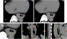
          A combination of four-dimensional computed tomography with 18F-fluorodeoxyglucose positron emission tomography (4D CT-FDG PET) was used to delineate gross tumor volume (GTV) in esophageal cancer (EC). Eighteen patients with EC were prospectively enrolled. Using 4D images taken during the respiratory cycle, the average CT image phase was fused with the average FDG PET phase in order to analyze the optimal standardized uptake values (SUV) or threshold. PET-based GTV (GTV PET) was determined with eight different threshold methods using the auto-contouring function on the PET workstation. The difference in volume ratio (VR) and conformality index (CI) between GTV PET and CT-based GTV (GTV CT) was investigated. The image sets via automatic co-registrations of 4D CT-FDG PET were available for 12 patients with 13 GTV CT values. The decision coefficient (R 2) of tumor length difference at the threshold levels of SUV 2.5, SUV 20% and SUV 25% were 0.79, 0.65 and 0.54, respectively. The mean volume of GTV CT was 29.41 ± 19.14 ml. The mean VR ranged from 0.30 to 1.48. The optimal VR of 0.98, close to 1, was at SUV 20% or SUV 2.5. The mean CI ranged from 0.28 to 0.58. The best CI was at SUV 20% (0.58) or SUV 2.5 (0.57). The auto-contouring function of the SUV threshold has the potential to assist in contouring the GTV. The SUV threshold setting of SUV 20% or SUV 2.5 achieves the optimal correlation of tumor length, VR, and CI using 4D-PET/CT images.

          Related collections

          Most cited references29

          • Record: found
          • Abstract: found
          • Article: not found

          Use of PET and PET/CT for radiation therapy planning: IAEA expert report 2006-2007.

          Positron Emission Tomography (PET) is a significant advance in cancer imaging with great potential for optimizing radiation therapy (RT) treatment planning and thereby improving outcomes for patients. The use of PET and PET/CT in RT planning was reviewed by an international panel. The International Atomic Energy Agency (IAEA) organized two synchronized and overlapping consultants' meetings with experts from different regions of the world in Vienna in July 2006. Nine experts and three IAEA staff evaluated the available data on the use of PET in RT planning, and considered practical methods for integrating it into routine practice. For RT planning, (18)F fluorodeoxyglucose (FDG) was the most valuable pharmaceutical. Numerous studies supported the routine use of FDG-PET for RT target volume determination in non-small cell lung cancer (NSCLC). There was also evidence for utility of PET in head and neck cancers, lymphoma and in esophageal cancers, with promising preliminary data in many other cancers. The best available approach employs integrated PET/CT images, acquired on a dual scanner in the radiotherapy treatment position after administration of tracer according to a standardized protocol, with careful optimization of images within the RT planning system and carefully considered rules for contouring tumor volumes. PET scans that are not recent or were acquired without proper patient positioning should be repeated for RT planning. PET will play an increasing valuable role in RT planning for a wide range of cancers. When requesting PET scans, physicians should be aware of their potential role in RT planning.
            Bookmark
            • Record: found
            • Abstract: found
            • Article: not found

            18F-FDG PET definition of gross tumor volume for radiotherapy of non-small cell lung cancer: is a single standardized uptake value threshold approach appropriate?

            PET with (18)F-FDG has been used in radiation treatment planning for non-small cell lung cancer (NSCLC). Thresholds of 15%-50% the maximum standardized uptake value (SUV(max)) have been used for gross tumor volume (GTV) delineation by PET (PET(GTV)), with 40% being the most commonly used value. Recent studies indicated that 15%-20% may be more appropriate. The purposes of this study were to determine which threshold generates the best volumetric match to GTV delineation by CT (CT(GTV)) for peripheral NSCLC and to determine whether that threshold can be generalized to tumors of various sizes. Data for patients who had peripheral NSCLC with well-defined borders on CT and SUV(max) of greater than 2.5 were reviewed. PET/CT datasets were reviewed, and a volume of interest was determined to represent the GTV. The CT(GTV) was delineated by using standard lung windows and reviewed by a radiation oncologist. The PET(GTV) was delineated automatically by use of various percentages of the SUV(max). The PET(GTV)-to-CT(GTV) ratios were compared at various thresholds, and a ratio of 1 was considered the best match, or the optimal threshold. Twenty peripheral NSCLCs with volumes easily defined on CT were evaluated. The SUV(max) (mean +/- SD) was 12 +/- 8, and the mean CT(GTV) was 198 cm(3) (97.5% confidence interval, 5-1,008). The SUV(max) were 16 +/- 5, 13 +/- 9, and 3.0 +/- 0.4 for tumors measuring greater than 5 cm, 3-5 cm, and less than 3 cm, respectively. The optimal thresholds (mean +/- SD) for the best match were 15% +/- 6% for tumors measuring greater than 5 cm, 24% +/- 9% for tumors measuring 3-5 cm, 42% +/- 2% for tumors measuring less than 3 cm, and 24% +/- 13% for all tumors. The PET(GTV) at the 40% and 20% thresholds underestimated the CT(GTV) for 16 of 20 and 14 of 20 lesions, respectively. The mean difference in the volumes (PET(GTV) minus CT(GTV) [PET(GTV) - CT(GTV)]) at the 20% threshold was 79 cm(3) (97.5% confidence interval, -922 to 178). The PET(GTV) at the 20% threshold overestimated the CT(GTV) for all 4 tumors measuring less than 3 cm and underestimated the CT(GTV) for all 6 tumors measuring greater than 5 cm. The CT(GTV) was inversely correlated with the PET(GTV) - CT(GTV) at the 20% threshold (R(2) = 0.90, P < 0.0001). The optimal threshold was inversely correlated with the CT(GTV) (R(2) = 0.79, P < 0.0001). No single threshold delineating the PET(GTV) provides accurate volume definition, compared with that provided by the CT(GTV), for the majority of NSCLCs. The strong correlation of the optimal threshold with the CT(GTV) warrants further investigation.
              Bookmark
              • Record: found
              • Abstract: found
              • Article: not found

              Systematic review of the staging performance of 18F-fluorodeoxyglucose positron emission tomography in esophageal cancer.

              Despite the increasing number of publications concerning (18)F-fluorodeoxyglucose positron emission tomography (FDG-PET) for staging of esophageal cancer and the increasing availability of this novel diagnostic modality, its exact role in preoperative staging of these tumors is still unknown. The aim of this study was to systematically review the literature regarding the diagnostic performance of FDG-PET in preoperative staging of patients with esophageal cancer, and to calculate summary estimates of its sensitivity and specificity. The databases of PubMed, Embase, and Cochrane were searched for relevant studies. Two reviewers independently assessed the methodological quality of each study. A meta-analysis of the reported sensitivity and specificity of each study was performed. Twelve studies met the inclusion criteria. The studies had several design deficiencies. Pooled sensitivity and specificity for the detection of locoregional metastases were 0.51 (95% CI, 0.34 to 0.69) and 0.84 (95% CI, 0.76 to 0.91), respectively. For distant metastases, pooled sensitivity and specificity were 0.67 (95% CI, 0.58 to 0.76) and 0.97 (95% CI, 0.90 to 1.0), respectively. FDG-PET showed moderate sensitivity and specificity for the detection of locoregional metastases, and reasonable sensitivity and specificity in detection of distant lymphatic and hematogenous metastases.
                Bookmark

                Author and article information

                Journal
                J Radiat Res
                J. Radiat. Res
                radres
                jrr
                Journal of radiation research
                Oxford University Press
                0449-3060
                1349-9157
                July 2012
                5 June 2012
                : 53
                : 4
                : 594-600
                Affiliations
                [1 ]Department of Radiation Oncology, China Medical University Hospital, Taichung, Taiwan
                [2 ]School of Medicine, Graduate Institute of Clinical Medical Science, China Medical University, Taichung, Taiwan
                [3 ]Department of Nuclear Medicine and PET Center, China Medical University Hospital, Taichung, Taiwan
                [4 ]Department of Biomedical Imaging and Radiological Science, China Medical University Hospital, Taichung, Taiwan
                [5 ]School of Medicine, China Medical University, Taichung, Taiwan
                [6 ]University of Texas MD Anderson Cancer Center, Houston, TX, USA
                Author notes
                [* ]Corresponding authors: Departments of Radiation Oncology and Nuclear Medicine, China Medical University Hospital, No. 2, Yuh-Der Road, Taichung 404, Taiwan; Tel: 886-4-22052121-7461; Fax: 886-4-22339372; Email: d4615@ 123456mail.cmuh.org.tw , d10040@ 123456mail.cmuh.org.tw
                [†]

                Chia-Hung Kao and Ji-An Liang contributed equally to this work.

                Article
                rrs009
                10.1093/jrr/rrs009
                3393356
                22843625
                bae910b9-6974-4522-a092-876040bf9971
                © The Author 2012. Published by Oxford University Press on behalf of The Japan Radiation Research Society and Japanese Society for Therapeutic Radiology and Oncology.

                This is an Open Access article distributed under the terms of the Creative Commons Attribution Non-Commercial License ( http://creativecommons.org/licenses/by-nc/3.0/), which permits unrestricted non-commercial use, distribution, and reproduction in any medium, provided the original work is properly cited.

                History
                : 1 November 2011
                : 5 March 2012
                : 16 March 2012
                Page count
                Pages: 28
                Categories
                Oncology

                Oncology & Radiotherapy
                fdg pet/ct,gross tumor volume,radiotherapy,esophageal cancer
                Oncology & Radiotherapy
                fdg pet/ct, gross tumor volume, radiotherapy, esophageal cancer

                Comments

                Comment on this article