19
views
0
recommends
+1 Recommend
0 collections
    0
    shares
      • Record: found
      • Abstract: found
      • Article: not found

      Cancer therapy related complications in the liver, pancreas, and biliary system: an imaging perspective

      review-article

      Read this article at

      ScienceOpenPublisherPMC
      Bookmark
          There is no author summary for this article yet. Authors can add summaries to their articles on ScienceOpen to make them more accessible to a non-specialist audience.

          Abstract

          Abstract

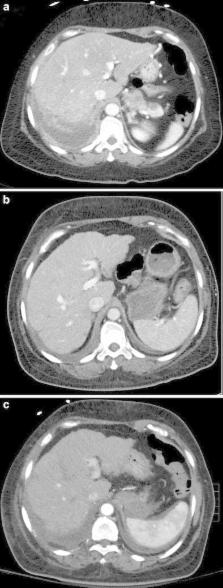
          Awareness of cancer therapy-induced toxicities is important for all clinicians treating patients with cancer. Cancer therapy has evolved to include classic cytotoxic agents in addition to newer options such as targeted agents and catheter-directed chemoembolisation. Several adverse affects can result from the wide array of treatments including effects on the liver, pancreas, and biliary system that can be visualised on imaging. These complications include sinusoidal obstruction syndrome, fatty liver, pseudocirrhosis, acute hepatitis, pancreatitis, pancreatic atrophy, cholecystitis, biliary sclerosis, and biliary stasis. Many of these toxicities are manageable and reversible with supportive therapies and/or cessation of cancer therapy. The objective of this review is to discuss the imaging findings associated with cancer therapy-induced toxicity of the liver, biliary system, and pancreas.

          Teaching Points

          Cancer therapy can have adverse effects on the hepatobiliary system and pancreas.

          Cancer therapy-induced toxicities can be visualised on imaging.

          Knowledge of imaging changes associated with cancer therapy complications can improve treatment.

          Related collections

          Most cited references65

          • Record: found
          • Abstract: found
          • Article: not found

          Toxic injury to hepatic sinusoids: sinusoidal obstruction syndrome (veno-occlusive disease).

          The term veno-occlusive disease of the liver refers to a form of toxic liver injury characterized clinically by the development of hepatomegaly, ascites, and jaundice, and histologically by diffuse damage in the centrilobular zone of the liver. The cardinal histologic features of this injury are marked sinusoidal fibrosis, necrosis of pericentral hepatocytes, and narrowing and eventual fibrosis of central veins. Recent studies suggest that the primary site of the toxic injury is sinusoidal endothelial cells, followed by a series of biologic processes that lead to circulatory compromise of centrilobular hepatocytes, fibrosis, and obstruction of liver blood flow. Thus we propose a more appropriate name for this form of liver injury--sinusoidal obstruction syndrome. This review encompasses historical perspectives, clinical manifestations of sinusoidal obstruction syndrome in the setting of hematopoietic cell transplantation, histologic features of centrilobular injury, and a discussion of the pathophysiology of sinusoidal injury, based on both animal and clinical investigations.
            Bookmark
            • Record: found
            • Abstract: found
            • Article: not found

            Drug-induced cholestasis.

            Recent progress in understanding the molecular mechanisms of bile formation and cholestasis have led to new insights into the pathogenesis of drug-induced cholestasis. This review summarizes their variable clinical presentations, examines the role of transport proteins in hepatic drug clearance and toxicity, and addresses the increasing importance of genetic determinants, as well as practical aspects of diagnosis and management.
              Bookmark
              • Record: found
              • Abstract: found
              • Article: not found

              Fatty liver: imaging patterns and pitfalls.

              Fat accumulation is one of the most common abnormalities of the liver depicted on cross-sectional images. Common patterns include diffuse fat accumulation, diffuse fat accumulation with focal sparing, and focal fat accumulation in an otherwise normal liver. Unusual patterns that may cause diagnostic confusion by mimicking neoplastic, inflammatory, or vascular conditions include multinodular and perivascular accumulation. All of these patterns involve the heterogeneous or nonuniform distribution of fat. To help prevent diagnostic errors and guide appropriate work-up and management, radiologists should be aware of the different patterns of fat accumulation in the liver, especially as they are depicted at ultrasonography, computed tomography, and magnetic resonance imaging. In addition, knowledge of the risk factors and the pathophysiologic, histologic, and epidemiologic features of fat accumulation may be useful for avoiding diagnostic pitfalls and planning an appropriate work-up in difficult cases. RSNA, 2006
                Bookmark

                Author and article information

                Contributors
                (714) 495-1175 , ngod@evms.edu
                Journal
                Insights Imaging
                Insights Imaging
                Insights into Imaging
                Springer Berlin Heidelberg (Berlin/Heidelberg )
                1869-4101
                6 October 2015
                6 October 2015
                December 2015
                : 6
                : 6
                : 665-677
                Affiliations
                [ ]Eastern Virginia Medical School, 700 W Olney Road, Norfolk, VA 23507 USA
                [ ]Department of Radiological Sciences, University of California, Irvine, 101 The City Drive South, Orange, CA 92868 USA
                Article
                436
                10.1007/s13244-015-0436-7
                4656242
                26443452
                bb7e5eb5-b8e6-418b-94a5-e29bb1e2915b
                © The Author(s) 2015

                Open Access This article is distributed under the terms of the Creative Commons Attribution 4.0 International License (http://creativecommons.org/licenses/by/4.0/), which permits unrestricted use, distribution, and reproduction in any medium, provided you give appropriate credit to the original author(s) and the source, provide a link to the Creative Commons license, and indicate if changes were made.

                History
                : 26 June 2015
                : 14 September 2015
                : 18 September 2015
                Categories
                Pictorial Review
                Custom metadata
                © The Author(s) 2015

                Radiology & Imaging
                cancer therapy,drug-associated adverse effects,hepatic toxicity,pancreatic toxicity,biliary toxicity

                Comments

                Comment on this article