20
views
0
recommends
+1 Recommend
2 collections
    0
    shares
      • Record: found
      • Abstract: found
      • Article: found
      Is Open Access

      Potential treatment of COVID-19 by inhibitors of human dihydroorotate dehydrogenase

      research-article
      1 , 2 , 1 , 2 , 3 ,
      Protein & Cell
      Higher Education Press

      Read this article at

      Bookmark
          There is no author summary for this article yet. Authors can add summaries to their articles on ScienceOpen to make them more accessible to a non-specialist audience.

          Abstract

          The ongoing pandemic of severe acute respiratory syndrome coronavirus 2 (SARS-CoV-2), referred to as coronavirus disease 2019 (COVID-19), has caused over 13 million infections and over 560,000 deaths worldwide (https://www.who.int/emergencies/diseases/novel-coronavirus-2019/situation-reports), posing a significant threat to globally public health and economics. At present, no efficacious antiviral drugs and vaccines have been approved for the prophylaxis or treatment of COVID-19. Tremendous efforts have been made to develop drug and vaccine against SARS-CoV-2. The main protease (Mpro, also called 3CLpro) is an attractive drug target among coronaviruses, and several potent inhibitors of the SARS-CoV-2 3CLpro together with their crystal structures in complex with the protease have been reported (Dai et al., 2020; Jin et al., 2020; Zhang et al., 2020). While the viral RNA-dependent RNA polymerases (RdRp) are well-known broad-spectrum antiviral drug targets, the cryo-electron microscopy structures of the SARS-CoV-2 RdRp and its complex with remdesivir, a promising antiviral candidate developed by Gilead Sciences, validated the efficient inhibition of the viral RNA replication by remdesivir and provided a rational template for drug design to combat SARS-CoV-2 infections (Gao et al., 2020; Wang et al., 2020; Yin et al., 2020). In addition, the trimeric spike protein on the surface of SARS-CoV-2 plays a pivotal role during the viral entry by binding to the peptidase domain of angiotensin-converting enzyme 2 (ACE2), a host cell receptor (Yan et al., 2020). It has been revealed that not only the receptor binding domain which is recognized by ACE2 but also the N-terminal domain of the SARS-CoV-2 spike protein is targeting sites for therapeutic monoclonal antibodies (Chi et al., 2020). Accordingly, both the inhibitors of 3CLpro or RdRp and the antibodies targeting the spike protein provide potential candidates for development of the direct-acting antiviral (DAA) drugs for the treatment of COVID-19. In addition to DAA drugs, host-targeting antiviral (HTA) agents, targeting host proteins required for the viral infection and replication, have advantages in overcoming drug resistance and combating a broad spectrum of viruses including the newly emerging virus (Ji and Li, 2020). Maraviroc, an antagonist of chemokine receptor type 5 for HIV treatment, presents a typical HTA drug. In a remarkable study published in this journal, Xiong et al. reported novel and potent inhibitors of human dihydroorotate dehydrogenase (DHODH) as broad-spectrum antiviral agents against RNA viruses including SARS-CoV-2 (Xiong et al., 2020). Pyrimidines serve as crucial building blocks for the biosynthesis of DNA, RNA, phospholipids, and glycoproteins, which is essential for the cell survival as well as proliferation (Loffler et al., 2005). Human DHODH belongs to the class 2 DHODH family and is a flavin-dependent mitochondrial enzyme catalyzing the oxidation of dihydroorotate to orotate, the fourth step also a rate limiting step in the de novo biosynthesis of pyrimidine-based nucleotides (Reis et al., 2017) (Fig. 1A). By consequence, DHODH is an attractive therapeutic target for multiple diseases including cancer and autoimmune diseases (Lolli et al., 2018; Boschi et al., 2019; Madak et al., 2019). Leflunomide and its metabolite teriflunomide, and brequinar are well-known DHODH inhibitors and were evaluated in clinical trials (Lolli et al., 2018). Leflunomide was approved for the therapy of rheumatoid arthritis many years ago (Herrmann et al., 2000).
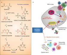
 Figure 1 DHODH in the de novo pyrimidine biosynthesis pathway and dual action of DHODHi. (A) DHODH catalyzes the fourth step in the de novo pyrimidine biosynthesis pathway. (B) DHODH inhibitors (DHODHi) are broad-spectrum antivirals against RNA viruses with the dual action of inhibiting viral genome replication and regulating the immune system With a computer-aided hit discovery and optimization strategy, Xiong et al. identified two novel and potent inhibitors of DHODH with a thiazole scaffold, S312 and S416 (Diao et al., 2012; Li et al., 2015; Zhu et al., 2015). The IC50s of these two compounds against human DHODH were 29.2 and 7.5 nmol/L, respectively, a >10-fold increase in activity relative to the FDA-approved teriflunomide (IC50 = 307.1 nmol/L). The X-ray crystal structure of DHODH in complex with S416 also revealed the binding mode of two inhibitors at the ubiquinone-binding site of the enzyme. Moreover, two inhibitors exhibited significant antiviral activities against influenza A (H1N1, H3N2 and H9N2), Zika, Ebola, and SARS-CoV-2 in cells infected with various tested viruses, demonstrating that DHODH inhibitors possess broad-spectrum antiviral activity by interfering the de novo pyrimidine synthesis pathway. Low toxicities of the inhibitors suggest that the reduced production of pyrimidine restricts virus replication but not cell growth. Most notably, the EC50 of S416 against the viral replication in the cells infected with SARS-CoV-2 at MOI of 0.05 is 17 nmol/L, and the resulting selectivity index (SI = CC50/EC50) reaches 10 505.88. It is much more potent than that of teriflunomide or brequinar and is also by far the most effective inhibitor against SARS-CoV-2 in cells. Another striking feature of this work is that S312 exhibited in vivo anti-influenza efficacy equivalent to that of oseltamivir, a marketed drug for the treatment of influenza. S312 at a dose of 5 mg/kg was also able to rescue all the influenza-infected mice from body weight loss and death. By contrast, previous studies often showed that inhibitors of either DHODH or the de novo pyrimidine biosynthesis pathway were ineffective against infection in animal models. In addition, the combination administration of S312 and oseltamivir resulted in 100% protection of the infected mice, superior to the single use of S312 or oseltamivir. S312 was also effective in the mice infected with an oseltamivir-resistant virus and had a remarkable advantage over oseltamivir to treat the late phase of the infectious disease. These results together demonstrated the feasibility of DHODH inhibitors used as efficacious antivirals as well as the combination of the DHODH inhibitor with DAA to overcome drug resistance. As leflunomide and teriflunomide are used to treat autoimmune diseases such as rheumatoid arthritis and multiple sclerosis by regulating lymphocytes and the release of cytokines and chemokines, it is reasonable to conjecture that S312 and S416 would have the similar efficacy too. As anticipated, the combination use of S312 and oseltamivir significantly reduced the levels of IL-6, MCP-1, IL-5, KC/GRO (CXCL1), IL-2, IFN-γ, IP-10, IL-9, TNF-α, GM-CSF, EPO, IL-12p70, MIP-3α, and IL-17A/F in the animal model. Therefore, the DHODH inhibitors not only inhibit the viral replication but also have regulatory roles in cytokine/chemokine production. Cytokine storm frequently occurred with patients suffered from virus infections such as SARS-CoV and SARS-CoV-2, antiviral treatment alone is thereby not enough and should be combined with appropriate anti-inflammatory treatment. The DHODH inhibitors provide the ideal candidate to take both into consideration. Taken together, this elegant work uncovers that DHODH is an attractive host target for developing broad-spectrum antivirals which achieve the efficacy through dual mechanism of action of antiviral and immuno-regulation (Fig. 1B), providing more therapeutic options in response to COVID-19 as well as other emergent RNA virus infections. In the present situation, S312 and S416, two potent inhibitors of DHODH with favorable drug-likeness and pharmacokinetic profiles, serve as right HTAs for further evaluation of therapeutic potential in COVID-19 treatment. Meanwhile, as a new concept for the treatment of COVID-19, the clinical trial of leflunomide has been initiated in England and founded by LifeArc (DEFEAT-COVID study) (https://www.lifearc.org/funding/covid-19-funding/).

          Related collections

          Most cited references11

          • Record: found
          • Abstract: found
          • Article: found
          Is Open Access

          Remdesivir and chloroquine effectively inhibit the recently emerged novel coronavirus (2019-nCoV) in vitro

          Dear Editor, In December 2019, a novel pneumonia caused by a previously unknown pathogen emerged in Wuhan, a city of 11 million people in central China. The initial cases were linked to exposures in a seafood market in Wuhan. 1 As of January 27, 2020, the Chinese authorities reported 2835 confirmed cases in mainland China, including 81 deaths. Additionally, 19 confirmed cases were identified in Hong Kong, Macao and Taiwan, and 39 imported cases were identified in Thailand, Japan, South Korea, United States, Vietnam, Singapore, Nepal, France, Australia and Canada. The pathogen was soon identified as a novel coronavirus (2019-nCoV), which is closely related to sever acute respiratory syndrome CoV (SARS-CoV). 2 Currently, there is no specific treatment against the new virus. Therefore, identifying effective antiviral agents to combat the disease is urgently needed. An efficient approach to drug discovery is to test whether the existing antiviral drugs are effective in treating related viral infections. The 2019-nCoV belongs to Betacoronavirus which also contains SARS-CoV and Middle East respiratory syndrome CoV (MERS-CoV). Several drugs, such as ribavirin, interferon, lopinavir-ritonavir, corticosteroids, have been used in patients with SARS or MERS, although the efficacy of some drugs remains controversial. 3 In this study, we evaluated the antiviral efficiency of five FAD-approved drugs including ribavirin, penciclovir, nitazoxanide, nafamostat, chloroquine and two well-known broad-spectrum antiviral drugs remdesivir (GS-5734) and favipiravir (T-705) against a clinical isolate of 2019-nCoV in vitro. Standard assays were carried out to measure the effects of these compounds on the cytotoxicity, virus yield and infection rates of 2019-nCoVs. Firstly, the cytotoxicity of the candidate compounds in Vero E6 cells (ATCC-1586) was determined by the CCK8 assay. Then, Vero E6 cells were infected with nCoV-2019BetaCoV/Wuhan/WIV04/2019 2 at a multiplicity of infection (MOI) of 0.05 in the presence of varying concentrations of the test drugs. DMSO was used in the controls. Efficacies were evaluated by quantification of viral copy numbers in the cell supernatant via quantitative real-time RT-PCR (qRT-PCR) and confirmed with visualization of virus nucleoprotein (NP) expression through immunofluorescence microscopy at 48 h post infection (p.i.) (cytopathic effect was not obvious at this time point of infection). Among the seven tested drugs, high concentrations of three nucleoside analogs including ribavirin (half-maximal effective concentration (EC50) = 109.50 μM, half-cytotoxic concentration (CC50) > 400 μM, selectivity index (SI) > 3.65), penciclovir (EC50 = 95.96 μM, CC50 > 400 μM, SI > 4.17) and favipiravir (EC50 = 61.88 μM, CC50 > 400 μM, SI > 6.46) were required to reduce the viral infection (Fig. 1a and Supplementary information, Fig. S1). However, favipiravir has been shown to be 100% effective in protecting mice against Ebola virus challenge, although its EC50 value in Vero E6 cells was as high as 67 μM, 4 suggesting further in vivo studies are recommended to evaluate this antiviral nucleoside. Nafamostat, a potent inhibitor of MERS-CoV, which prevents membrane fusion, was inhibitive against the 2019-nCoV infection (EC50 = 22.50 μM, CC50 > 100 μM, SI > 4.44). Nitazoxanide, a commercial antiprotozoal agent with an antiviral potential against a broad range of viruses including human and animal coronaviruses, inhibited the 2019-nCoV at a low-micromolar concentration (EC50 = 2.12 μM; CC50 > 35.53 μM; SI > 16.76). Further in vivo evaluation of this drug against 2019-nCoV infection is recommended. Notably, two compounds remdesivir (EC50 = 0.77 μM; CC50 > 100 μM; SI > 129.87) and chloroquine (EC50 = 1.13 μM; CC50 > 100 μM, SI > 88.50) potently blocked virus infection at low-micromolar concentration and showed high SI (Fig. 1a, b). Fig. 1 The antiviral activities of the test drugs against 2019-nCoV in vitro. a Vero E6 cells were infected with 2019-nCoV at an MOI of 0.05 in the treatment of different doses of the indicated antivirals for 48 h. The viral yield in the cell supernatant was then quantified by qRT-PCR. Cytotoxicity of these drugs to Vero E6 cells was measured by CCK-8 assays. The left and right Y-axis of the graphs represent mean % inhibition of virus yield and cytotoxicity of the drugs, respectively. The experiments were done in triplicates. b Immunofluorescence microscopy of virus infection upon treatment of remdesivir and chloroquine. Virus infection and drug treatment were performed as mentioned above. At 48 h p.i., the infected cells were fixed, and then probed with rabbit sera against the NP of a bat SARS-related CoV 2 as the primary antibody and Alexa 488-labeled goat anti-rabbit IgG (1:500; Abcam) as the secondary antibody, respectively. The nuclei were stained with Hoechst dye. Bars, 100 μm. c and d Time-of-addition experiment of remdesivir and chloroquine. For “Full-time” treatment, Vero E6 cells were pre-treated with the drugs for 1 h, and virus was then added to allow attachment for 2 h. Afterwards, the virus–drug mixture was removed, and the cells were cultured with drug-containing medium until the end of the experiment. For “Entry” treatment, the drugs were added to the cells for 1 h before viral attachment, and at 2 h p.i., the virus–drug mixture was replaced with fresh culture medium and maintained till the end of the experiment. For “Post-entry” experiment, drugs were added at 2 h p.i., and maintained until the end of the experiment. For all the experimental groups, cells were infected with 2019-nCoV at an MOI of 0.05, and virus yield in the infected cell supernatants was quantified by qRT-PCR c and NP expression in infected cells was analyzed by Western blot d at 14 h p.i. Remdesivir has been recently recognized as a promising antiviral drug against a wide array of RNA viruses (including SARS/MERS-CoV 5 ) infection in cultured cells, mice and nonhuman primate (NHP) models. It is currently under clinical development for the treatment of Ebola virus infection. 6 Remdesivir is an adenosine analogue, which incorporates into nascent viral RNA chains and results in pre-mature termination. 7 Our time-of-addition assay showed remdesivir functioned at a stage post virus entry (Fig. 1c, d), which is in agreement with its putative anti-viral mechanism as a nucleotide analogue. Warren et al. showed that in NHP model, intravenous administration of 10 mg/kg dose of remdesivir resulted in concomitant persistent levels of its active form in the blood (10 μM) and conferred 100% protection against Ebola virus infection. 7 Our data showed that EC90 value of remdesivir against 2019-nCoV in Vero E6 cells was 1.76 μM, suggesting its working concentration is likely to be achieved in NHP. Our preliminary data (Supplementary information, Fig. S2) showed that remdesivir also inhibited virus infection efficiently in a human cell line (human liver cancer Huh-7 cells), which is sensitive to 2019-nCoV. 2 Chloroquine, a widely-used anti-malarial and autoimmune disease drug, has recently been reported as a potential broad-spectrum antiviral drug. 8,9 Chloroquine is known to block virus infection by increasing endosomal pH required for virus/cell fusion, as well as interfering with the glycosylation of cellular receptors of SARS-CoV. 10 Our time-of-addition assay demonstrated that chloroquine functioned at both entry, and at post-entry stages of the 2019-nCoV infection in Vero E6 cells (Fig. 1c, d). Besides its antiviral activity, chloroquine has an immune-modulating activity, which may synergistically enhance its antiviral effect in vivo. Chloroquine is widely distributed in the whole body, including lung, after oral administration. The EC90 value of chloroquine against the 2019-nCoV in Vero E6 cells was 6.90 μM, which can be clinically achievable as demonstrated in the plasma of rheumatoid arthritis patients who received 500 mg administration. 11 Chloroquine is a cheap and a safe drug that has been used for more than 70 years and, therefore, it is potentially clinically applicable against the 2019-nCoV. Our findings reveal that remdesivir and chloroquine are highly effective in the control of 2019-nCoV infection in vitro. Since these compounds have been used in human patients with a safety track record and shown to be effective against various ailments, we suggest that they should be assessed in human patients suffering from the novel coronavirus disease. Supplementary information Supplementary information, Materials and Figures
            Bookmark
            • Record: found
            • Abstract: found
            • Article: found
            Is Open Access

            Structural basis for the recognition of SARS-CoV-2 by full-length human ACE2

            How SARS-CoV-2 binds to human cells Scientists are racing to learn the secrets of severe acute respiratory syndrome–coronavirus 2 (SARS-CoV-2), which is the cause of the pandemic disease COVID-19. The first step in viral entry is the binding of the viral trimeric spike protein to the human receptor angiotensin-converting enzyme 2 (ACE2). Yan et al. present the structure of human ACE2 in complex with a membrane protein that it chaperones, B0AT1. In the context of this complex, ACE2 is a dimer. A further structure shows how the receptor binding domain of SARS-CoV-2 interacts with ACE2 and suggests that it is possible that two trimeric spike proteins bind to an ACE2 dimer. The structures provide a basis for the development of therapeutics targeting this crucial interaction. Science, this issue p. 1444
              Bookmark
              • Record: found
              • Abstract: found
              • Article: not found

              Structure of Mpro from COVID-19 virus and discovery of its inhibitors

              A new coronavirus, known as severe acute respiratory syndrome coronavirus 2 (SARS-CoV-2), is the aetiological agent responsible for the 2019-2020 viral pneumonia outbreak of coronavirus disease 2019 (COVID-19)1-4. Currently, there are no targeted therapeutic agents for the treatment of this disease, and effective treatment options remain very limited. Here we describe the results of a programme that aimed to rapidly discover lead compounds for clinical use, by combining structure-assisted drug design, virtual drug screening and high-throughput screening. This programme focused on identifying drug leads that target main protease (Mpro) of SARS-CoV-2: Mpro is a key enzyme of coronaviruses and has a pivotal role in mediating viral replication and transcription, making it an attractive drug target for SARS-CoV-25,6. We identified a mechanism-based inhibitor (N3) by computer-aided drug design, and then determined the crystal structure of Mpro of SARS-CoV-2 in complex with this compound. Through a combination of structure-based virtual and high-throughput screening, we assayed more than 10,000 compounds-including approved drugs, drug candidates in clinical trials and other pharmacologically active compounds-as inhibitors of Mpro. Six of these compounds inhibited Mpro, showing half-maximal inhibitory concentration values that ranged from 0.67 to 21.4 μM. One of these compounds (ebselen) also exhibited promising antiviral activity in cell-based assays. Our results demonstrate the efficacy of our screening strategy, which can lead to the rapid discovery of drug leads with clinical potential in response to new infectious diseases for which no specific drugs or vaccines are available.
                Bookmark

                Author and article information

                Contributors
                hljiang@simm.ac.cn
                Journal
                Protein Cell
                Protein Cell
                Protein & Cell
                Higher Education Press (Beijing )
                1674-800X
                1674-8018
                6 August 2020
                6 August 2020
                : 1-4
                Affiliations
                [1 ]GRID grid.9227.e, ISNI 0000000119573309, CAS Key Laboratory of Receptor Research, Shanghai Institute of Materia Medica, , Chinese Academy of Sciences, ; Shanghai, 201203 China
                [2 ]GRID grid.410726.6, ISNI 0000 0004 1797 8419, University of Chinese Academy of Sciences, ; Beijing, 100049 China
                [3 ]GRID grid.440637.2, ISNI 0000 0004 4657 8879, Shanghai Institute for Advanced Immunochemical Studies and School of Life Science and Technology, , ShanghaiTech University, ; Shanghai, 201210 China
                Article
                769
                10.1007/s13238-020-00769-9
                7406694
                32761523
                bc1adf5c-b662-4803-9903-cb0d47379cac
                © The Author(s) 2020

                Open AccessThis article is licensed under a Creative Commons Attribution 4.0 International License, which permits use, sharing, adaptation, distribution and reproduction in any medium or format, as long as you give appropriate credit to the original author(s) and the source, provide a link to the Creative Commons licence, and indicate if changes were made. The images or other third party material in this article are included in the article's Creative Commons licence, unless indicated otherwise in a credit line to the material. If material is not included in the article's Creative Commons licence and your intended use is not permitted by statutory regulation or exceeds the permitted use, you will need to obtain permission directly from the copyright holder. To view a copy of this licence, visit http://creativecommons.org/licenses/by/4.0/.

                History
                Categories
                Highlight

                Comments

                Comment on this article