55
views
0
recommends
+1 Recommend
0 collections
    0
    shares
      • Record: found
      • Abstract: found
      • Article: found
      Is Open Access

      Stable Expression of Human Muscle-Specific Kinase in HEp-2 M4 Cells for Automatic Immunofluorescence Diagnostics of Myasthenia Gravis

      research-article

      Read this article at

      Bookmark
          There is no author summary for this article yet. Authors can add summaries to their articles on ScienceOpen to make them more accessible to a non-specialist audience.

          Abstract

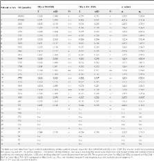
          Muscle-specific kinase (MuSK) belongs to the nicotinic acetylcholine receptor complex which is targeted by pathogenic autoantibodies causing Myasthenia gravis. While up to 95% of patients with generalized Myasthenia gravis were shown to be positive for acetylcholine receptor-specific autoantibodies, up to 70% of the remaining patients develop autoantibodies against MuSK. Discrimination of the autoantibody specificity is important for therapy of Myasthenia gravis. Recently, the new automatic fluorescence assessment platform AKLIDES has been developed for immunofluorescence-based diagnostics of autoimmune diseases. In order to establish an AKLIDES procedure for the detection of MuSK-specific autoantibodies (anti-MuSK), we developed a recombinant HEp-2 cell clone expressing the human MuSK cDNA. Here we show at the mRNA and protein level that the cell clone HEp-2 M4 stably expresses human MuSK. We provide evidence for a localization of MuSK at the cell membrane. Using cell clone HEp-2 M4 on the AKLIDES system, we investigated 34 patient sera that were previously tested anti-MuSK positive by radioimmunoassay as positive controls. As negative controls, we tested 29 acetylcholine receptor-positive but MuSK-negative patient sera, 30 amytrophic lateral sclerosis (ALS) patient sera and 45 blood donors. HEp-2 M4 cells revealed a high specificity for the detection of MuSK autoantibodies from 25 patient sera assessed by a specific pattern on HEp-2 M4 cells. By using appropriate cell culture additives, the fraction of cells stained positive with anti-MuSK containing sera can be increased from 2–16% to 10–48%, depending on the serum. In conclusion, we provide data showing that the novel recombinant cell line HEp-2 M4 can be used to screen for anti-MuSK with the automatic AKLIDES system.

          Related collections

          Most cited references45

          • Record: found
          • Abstract: found
          • Article: not found

          Methylated DNA and MeCP2 recruit histone deacetylase to repress transcription.

          CpG methylation in vertebrates correlates with alterations in chromatin structure and gene silencing. Differences in DNA-methylation status are associated with imprinting phenomena and carcinogenesis. In Xenopus laevis oocytes, DNA methylation dominantly silences transcription through the assembly of a repressive nucleosomal array. Methylated DNA assembled into chromatin binds the transcriptional repressor MeCP2 which cofractionates with Sin3 and histone deacetylase. Silencing conferred by MeCP2 and methylated DNA can be relieved by inhibition of histone deacetylase, facilitating the remodelling of chromatin and transcriptional activation. These results establish a direct causal relationship between DNA methylation-dependent transcriptional silencing and the modification of chromatin.
            Bookmark
            • Record: found
            • Abstract: found
            • Article: not found

            Myasthenia gravis: past, present, and future.

            Myasthenia gravis (MG) is an autoimmune syndrome caused by the failure of neuromuscular transmission, which results from the binding of autoantibodies to proteins involved in signaling at the neuromuscular junction (NMJ). These proteins include the nicotinic AChR or, less frequently, a muscle-specific tyrosine kinase (MuSK) involved in AChR clustering. Much is known about the mechanisms that maintain self tolerance and modulate anti-AChR Ab synthesis, AChR clustering, and AChR function as well as those that cause neuromuscular transmission failure upon Ab binding. This insight has led to the development of improved diagnostic methods and to the design of specific immunosuppressive or immunomodulatory treatments.
              Bookmark
              • Record: found
              • Abstract: found
              • Article: not found

              Analysis of cell cycle by flow cytometry.

              Described are four widely used procedures to analyze the cell cycle by flow cytometry. The first two are based on univariate analysis of cellular DNA content following cell staining with either propidium iodide (PI) or 4',6'-diamidino-2-phenylindole (DAPI) and deconvolution of the cellular DNA content frequency histograms. This approach reveals distribution of cells in three major phases of the cycle (G1 vs S vs G2/M) and makes it possible to detect apoptotic cells with fractional DNA content. The third approach is based on the bivariate analysis of DNA content and proliferation-associated proteins. The expression of cyclin D, cyclin E, cyclin A, or cyclin B1 vs DNA content is presented as an example. This approach allows one to distinguish, for example, G0 from G1 cells, identify mitotic cells, or relate expression of other intracellular proteins to the cell cycle position. The fourth procedure relies on the detection of 5'-bromo-2'-deoxyuridine (BrdU) incorporation to label the DNA-replicating cells.
                Bookmark

                Author and article information

                Contributors
                Role: Editor
                Journal
                PLoS One
                PLoS ONE
                plos
                plosone
                PLoS ONE
                Public Library of Science (San Francisco, USA )
                1932-6203
                2014
                9 January 2014
                : 9
                : 1
                : e83924
                Affiliations
                [1 ]Faculty of Science, Brandenburg University of Technology Cottbus-Senftenberg, Senftenberg, Germany
                [2 ]GA Generic Assays GmbH, Dahlewitz/Berlin, Germany
                [3 ]Institute of Immunology, Technical University Dresden, Dresden, Germany
                Istanbul University, Turkey
                Author notes

                Competing Interests: Dirk Roggenbuck is a shareholder of GA Generic Assays GmbH and Medipan GmbH. Ilka Knütter and Rico Hiemann are employees of GA Generic Assays GmbH. This does not alter the authors' adherence to all the PLOS ONE policies on sharing data and material. The remaining authors declare that they have no competing financial or other interests.

                Conceived and designed the experiments: SG DR KC IK J-HK. Performed the experiments: SG SP IK NR RH. Analyzed the data: SG IK NR RH. Contributed reagents/materials/analysis tools: KC J-HK SG IK SP DR. Wrote the paper: SG J-HK DR CK. Experimental design: J-HK DR IK SG. Software adaption of used for analysis: RH. Stable clone establishment: SG. Immunofluorescence optmization: SG IK. Sera tests: SG IK NR SP. Providing sera: KC ND. Plasmid constructs: SP.

                Article
                PONE-D-13-32944
                10.1371/journal.pone.0083924
                3886972
                bd956904-e6c7-420e-92b6-056c73253d8b
                Copyright @ 2014

                This is an open-access article distributed under the terms of the Creative Commons Attribution License, which permits unrestricted use, distribution, and reproduction in any medium, provided the original author and source are credited.

                History
                : 9 August 2013
                : 11 November 2013
                Page count
                Pages: 9
                Funding
                This work was partially funded by ZIM project KF2088004AJ9. The funders had no role in study design, data collection and analysis, decision to publish, or preparation of the manuscript.
                Categories
                Research Article
                Biology
                Biotechnology
                Genetic Engineering
                Genetically Modified Organisms
                Immunology
                Antigen Processing and Recognition
                Molecular Cell Biology
                Gene Expression
                DNA modification
                Protein Translation
                Medicine
                Clinical Immunology
                Autoimmune Diseases
                Myasthenia Gravis
                Diagnostic Medicine

                Uncategorized
                Uncategorized

                Comments

                Comment on this article