7
views
0
recommends
+1 Recommend
0 collections
    0
    shares
      • Record: found
      • Abstract: found
      • Article: found
      Is Open Access

      Migration from full‐head mask to “open‐face” mask for immobilization of patients with head and neck cancer

      research-article

      Read this article at
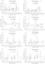
      Bookmark
          There is no author summary for this article yet. Authors can add summaries to their articles on ScienceOpen to make them more accessible to a non-specialist audience.

          Abstract

          To provide an alternative device for immobilization of the head while easing claustrophobia and improving comfort, an “open‐face” thermoplastic mask was evaluated using video‐based optical surface imaging (OSI) and kilovoltage (kV) X‐ray radiography. A three‐point thermoplastic head mask with a precut opening and reinforced strips was developed. After molding, it provided sufficient visible facial area as the region of interest for OSI. Using real‐time OSI, the head motion of ten volunteers in the new mask was evaluated during mask locking and 15 minutes lying on the treatment couch. Using a nose mark with reference to room lasers, forced head movement in open‐face and full‐head masks (with a nose hole) was compared. Five patients with claustrophobia were immobilized with open‐face masks, set up using OSI and kV, and treated in 121 fractions, in which 61 fractions were monitored during treatment using real‐time OSI. With the open‐face mask, head motion was found to be 1.0 ± 0.6 mm and 0.4 ° ± 0.2 ° in volunteers during the experiment, and 0.8 ± 0.3 mm and 0.4 ° ± 0.2 ° in patients during treatment. These agree with patient motion calculated from pre‐/post‐treatment OSI and kV data using different anatomical landmarks. In volunteers, the head shift induced by mask‐locking was 2.3 ± 1.7 mm and 1.8 ° ± 0.6 ° , and the range of forced movements in the open‐face and full‐head masks were found to be similar. Most (80%) of the volunteers preferred the open‐face mask to the full‐head mask, while claustrophobic patients could only tolerate the open‐face mask. The open‐face mask is characterized for its immobilization capability and can immobilize patients sufficiently ( < 2 mm ) during radiotherapy. It provides a clinical solution to the immobilization of patients with head and neck (HN) cancer undergoing radiotherapy, and is particularly beneficial for claustrophobic patients. This new open‐face mask is readily adopted in radiotherapy clinic as a superior alternative to the standard full‐head mask.

          PACS numbers: 87.19.xj, 87.63.L‐, 87.59.‐e, 87.55.tg, 87.55.‐x

          Related collections

          Most cited references19

          • Record: found
          • Abstract: found
          • Article: not found

          Inter- and intrafraction patient positioning uncertainties for intracranial radiotherapy: a study of four frameless, thermoplastic mask-based immobilization strategies using daily cone-beam CT.

          To determine whether frameless thermoplastic mask-based immobilization is adequate for image-guided cranial radiosurgery. Cone-beam CT localization data from patients with intracranial tumors were studied using daily pre- and posttreatment scans. The systems studied were (1) Type-S IMRT (head only) mask (Civco) with head cushion; (2) Uni-Frame mask (Civco) with head cushion, coupled with a BlueBag body immobilizer (Medical Intelligence); (3) Type-S head and shoulder mask with head and shoulder cushion (Civco); (4) same as previous, coupled with a mouthpiece. The comparative metrics were translational shift magnitude and average rotation angle; systematic inter-, random inter-, and random intrafraction positioning error was computed. For strategies 1-4, respectively, the analysis for interfraction variability included data from 20, 9, 81, and 11 patients, whereas that for intrafraction variability included a subset of 7, 9, 16, and 8 patients. The results were compared for statistical significance using an analysis of variance test. Immobilization system 4 provided the best overall accuracy and stability. The mean interfraction translational shifts (± SD) were 2.3 (± 1.4), 2.2 (± 1.1), 2.7 (± 1.5), and 2.1 (± 1.0) mm whereas intrafraction motion was 1.1 (± 1.2), 1.1 (± 1.1), 0.7 (± 0.9), and 0.7 (± 0.8) mm for devices 1-4, respectively. No significant correlation between intrafraction motion and treatment time was evident, although intrafraction motion was not purely random. We find that all frameless thermoplastic mask systems studied are viable solutions for image-guided intracranial radiosurgery. With daily pretreatment corrections, symmetric PTV margins of 1 mm would likely be adequate if ideal radiation planning and targeting systems were available. Copyright © 2011 Elsevier Inc. All rights reserved.
            Bookmark
            • Record: found
            • Abstract: found
            • Article: not found

            Skin toxicity due to intensity-modulated radiotherapy for head-and-neck carcinoma.

            To investigate the cause of acute skin toxicity observed in the treatment of head-and-neck cancer with extended-field intensity-modulated radiotherapy (EF-IMRT). EF-IMRT was used to treat head-and-neck cancer, with the gross target volume receiving 70 Gy and the clinical target volume 60 Gy. A thermoplastic mask covering the head, neck, and shoulder was used for immobilization. Dosimetric studies were conducted to investigate the possible causes of the skin reactions, such as the bolus effect of the mask, the use of multiple tangential beams with IMRT plans, and the way in which the physicians contoured the lymph nodes. The dose-volume histograms of conventional opposed-lateral fields were compared with that of the multiple tangential EF-IMRT fields. IMRT plans with neck nodes contoured up to and including the skin surface were compared with plans that contoured the neck nodes 5 mm away from the skin surface. In addition, IMRT plans defining the skin as a sensitive structure were compared with plans that did not define the skin as a sensitive structure. All plans were created using an anthropomorphic Rando phantom, and the skin doses were measured with and without the mask. In each measurement, 6 thermoluminescent dosimeters (TLDs) were placed at the lateral and medial surfaces of the neck. For all four plans, the measured skin doses with the mask were consistently higher than those without the mask. The average dose increase was about 18% owing to the bolus effect of the mask. Multiple tangential fields used in IMRT plans contributed to an increase in skin dose by about 19% and 27%, with and without the mask, respectively. If the skin of the neck was contoured as a sensitive structure for dose optimization, the volume of skin that received >45 Gy was further reduced by about 20%. Five patients immobilized with head and shoulder masks were treated with EF-IMRT plans with the neck nodes carefully delineated away from the skin surface. The neck skin was identified as a sensitive structure for dose optimization. Grade 1 toxicity was observed in 3 patients, Grade 2 in 1 patient, and Grade 3 in 1 patient toward the end of treatment. Multiple factors contributed to the observed acute skin reaction for head-and-neck cancer patients treated with EF-IMRT. By taking into consideration the skin as a sensitive structure during inverse planning, it was possible to reduce the skin dose to a tolerable level without compromising tumor target coverage.
              Bookmark
              • Record: found
              • Abstract: found
              • Article: not found

              Motion monitoring for cranial frameless stereotactic radiosurgery using video-based three-dimensional optical surface imaging.

              To establish a new clinical procedure in frameless stereotactic radiosurgery (SRS) for patient setup verification at treatment couch angles as well as for head-motion monitoring during treatment using video-based optical surface imaging (OSI).
                Bookmark

                Author and article information

                Contributors
                lig2@mskcc.org
                Journal
                J Appl Clin Med Phys
                J Appl Clin Med Phys
                10.1002/(ISSN)1526-9914
                ACM2
                Journal of Applied Clinical Medical Physics
                John Wiley and Sons Inc. (Hoboken )
                1526-9914
                06 September 2013
                September 2013
                : 14
                : 5 ( doiID: 10.1002/acm2.2013.14.issue-5 )
                : 243-254
                Affiliations
                [ 1 ] Department of Medical Physics Memorial Sloan‐Kettering Cancer Center New York NY USA
                [ 2 ] Department of Radiation Oncology Memorial Sloan‐Kettering Cancer Center New York NY USA
                Author notes
                [*] [* ]Corresponding author: Guang Li, Department of Medical Physics, Memorial Sloan‐Kettering Cancer Center, 1275 York Ave., New York, NY 10065, USA; phone: (212) 639 2891; fax: (212) 717 3258; email: lig2@ 123456mskcc.org
                Article
                ACM20243
                10.1120/jacmp.v14i5.4400
                5714571
                24036878
                bde5b962-d104-42b5-a277-82ce6e842bfb
                © 2013 The Authors.

                This is an open access article under the terms of the Creative Commons Attribution License, which permits use, distribution and reproduction in any medium, provided the original work is properly cited.

                History
                : 04 February 2013
                : 15 May 2013
                Page count
                Figures: 5, Tables: 3, References: 19, Pages: 12, Words: 6072
                Categories
                Radiation Oncology Physics
                Radiation Oncology Physics
                Custom metadata
                2.0
                acm20243
                September 2013
                Converter:WILEY_ML3GV2_TO_NLMPMC version:5.2.5 mode:remove_FC converted:16.11.2017

                patient setup and immobilization,thermoplastic head mask,image‐guided radiotherapy,optical surface imaging,motion uncertainty

                Comments

                Comment on this article