3
views
0
recommends
+1 Recommend
0 collections
    0
    shares
      • Record: found
      • Abstract: found
      • Article: found
      Is Open Access

      Evaluation of demographic and rare clinical characteristics of patients with thoracic carcinoid tumor in Razi and Aria Hospitals of Rasht during 2006-2016

      research-article

      Read this article at

      Bookmark
          There is no author summary for this article yet. Authors can add summaries to their articles on ScienceOpen to make them more accessible to a non-specialist audience.

          Abstract

          Introduction:

          Carcinoid tumors are malignant neoplasms of neuroendocrine cells. This study tended to evaluate the demographic and rare clinical characteristics of patients with thoracic carcinoid tumor during 2006-2016 at Razi and Aria Hospitals in Rasht.

          Materials and Methods:

          The present study was performed on records of 43 patients with lung carcinoid tumors referred to Razi and Aria Hospitals of Rasht during 2006-2016. Information on age, gender, rare clinical symptoms, smoking history, diagnosis tools, treatment, and outcome were analyzed.

          Results:

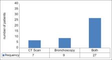
          Of 43 patients treated with definitive diagnosis of carcinoid tumor pathology, 31 patients had typic carcinoid tumor and 12 patients with atypic carcinoma (mean age 43.14 ± 15.16 years). The most common clinical symptom was cough and hemoptysis. Two cases presented with cushing syndrome, The most common diagnostic method in this study was simultaneous use of both CT scans and bronchoscopy. In 95.3% of cases, the tumor was pulmonary and in 4.7% of cases, it was extrapulmonary. Right lower lobe was the most common site of tumors and most of the surgeries used were lobectomy.

          Conclusion:

          This study showed that the most common clinical sign of thoracic carcinoid is cough and the tumor is pulmonary in 95.3% of cases. Right lower lobe was the most common site of tumors and most of the surgeries used were lobectomy. outcome was good.

          Related collections

          Most cited references30

          • Record: found
          • Abstract: found
          • Article: not found

          An analysis of 8305 cases of carcinoid tumors.

          Carcinoid tumors are unusual and most reports are anecdotal or limited in number. A series of 2837 cases was published in 1975. No recent large series is available. The authors evaluated 5468 cases identified by the Surveillance, Epidemiology, and End Results (SEER) program of the National Cancer Institute (NCI) from 1973 to 1991 together with 2837 carcinoid cases previously registered by 2 earlier NCI programs. To the authors' knowledge, the 8305 carcinoid tumors analyzed represent the largest current epidemiology series to date. The most frequent sites for carcinoids were the gastrointestinal (GI) tract (73.7%) and the bronchopulmonary system (25.1%). Within the GI tract, most occurred in the small bowel (28.7%), appendix (18.9%), and rectum (12.6%). For all sites, age-adjusted incidence rates were highest in African American males (2.12 per 100,000 population per year). Associated noncarcinoid tumors were frequent in conjunction with small intestinal (16.6%), appendiceal (14.6%), and colonic carcinoids (13.1%). The highest percentage of nonlocalized lesions were noted for pancreatic (76.1%), colonic (71.2%), and small intestinal carcinoids (70.7%) and this corresponded to their poor 5-year survival rates (34.1%, 41.6%, and 55.4%, respectively). The best 5-year survival rates were recorded for appendiceal (85.9%), bronchopulmonary (76.6%), and rectal carcinoids (72.2%). These exhibited invasive growth or metastatic spread in only 35.4%, 27.2%, and 14.2% of cases, respectively. Carcinoids appear to have increased in incidence in the past 20 years. In part, this may be due to different surgical rules of the various registries, improved diagnostic technology, and increased awareness. A cumulative analysis of all types of carcinoid tumors in the SEER group indicates that in 45.3% metastases are already evident at the time of diagnosis. The overall 5-year survival rate of all carcinoid tumors regardless of site was 50.4% +/- 6.4%.
            Bookmark
            • Record: found
            • Abstract: found
            • Article: not found

            Pulmonary atypical carcinoid: predictors of survival in 106 cases.

            Pulmonary neuroendocrine tumors (NE) include a spectrum of tumors from typical carcinoid (TC) to atypical carcinoid (AC), large cell neuroendocrine carcinoma (LCNEC), and small cell carcinoma (SCLC). Little is known about prognostic predictors for AC because of its rarity. Survival analysis was performed on 106 ACs with clinical follow-up from the AFIP and the Pathology Panel of the International Association for the Study of Lung Cancer (IASLC). The tumors fulfilled the 1999 WHO/IASLC criteria for AC of a NE tumor with a mitotic rate of 2 to 10 per 2 mm(2) of viable tumor or coagulative necrosis. Multiple clinical and histologic features were analyzed by Kaplan-Meier and Cox regression analysis. Of the clinical features, higher stage (P = .003) and a tumor size of 3.5 cm or greater (P = .003) were associated with a worse prognosis. Features that were histologically unfavorable by univariate analysis were mitotic rate (P =.002), pleomorphism (P = .018), and aerogenous spread (P =.007). Histologically favorable features by univariate analysis were the presence of palisading (P = .008), papillary (P = .039), pseudoglandular (P =.026), and rosette (P = .022) patterns. Female gender showed a trend toward a poorer prognosis (P =.085) and was included in the multivariate model. Multivariate analysis stratified for stage showed mitoses (P<.001), a tumor size of 3.5 cm or greater (P =.017), and female gender (P =.012) to be the only negative independent predictors of prognosis and the presence of rosettes (P = .016) to be the only independent positive predictor. We further divided the AC into subgroups of low (2 to 5 mitoses/2 mm(2)) and high (6 to 10 mitoses/2 mm(2)) mitotic rate and compared the survival with TC and with LCNEC. Within the category of AC, the patients with a higher mitotic rate had a significantly worse survival than those with a lower mitotic rate (P<.001) stratified for stage. Five- and 10-year survival rates for AC (61% and 35%, respectively) stratified for stage were significantly worse than for TC and better than that for LCNEC and SCLC. Chemotherapy or radiation therapy was given in 12 of 52 and 14 of 52 cases, respectively, but the data were insufficient to evaluate tumor response. We conclude that AC is an aggressive neuroendocrine neoplasm with survival intermediate between TC and LCNEC and SCLC. Higher mitotic rate, tumor size of 3.5 cm or greater, female gender, and presence of rosettes are the only independent predictors of survival. Surgical resection remains the treatment of choice, and the role of chemotherapy and radiation therapy remains to be proven.
              Bookmark
              • Record: found
              • Abstract: found
              • Article: not found

              The gastroenteropancreatic neuroendocrine cell system and its tumors: the WHO classification.

              Although well established in medical terminology, the term carcinoid is no longer adequate to cover the entire morphological and biological spectrum of neoplasms of the disseminated neuroendocrine cell system. Therefore, instead of carcinoid, the WHO classification published in 2000 uses the general terms neuroendocrine tumor and neuroendocrine carcinoma. In this review a classification of gastroenteropancreatic neuroendocrine tumors based on the WHO criteria is described. We also classify and comment on the most important tumor entities. On the basis of localization and of various morphological and biological criteria, we distinguish between benign neuroendocrine tumors, tumors with uncertain malignant potential, and tumors showing low-grade and high-grade malignancy.
                Bookmark

                Author and article information

                Journal
                J Family Med Prim Care
                J Family Med Prim Care
                JFMPC
                J Family Med Prim Care
                Journal of Family Medicine and Primary Care
                Wolters Kluwer - Medknow (India )
                2249-4863
                2278-7135
                September 2022
                14 October 2022
                : 11
                : 9
                : 5437-5441
                Affiliations
                [1] Inflammatory Lung Diseases Research Center, Department of Internal Medicine, Razi Hospital, School of Medicine, Guilan University of Medical Sciences, Rasht, Iran
                Author notes
                Address for correspondence: Dr. Mohammad Reza Asgary, Inflammatory Lung Diseases Research Center, Department of Internal Medicine, Razi Hospital, School of Medicine, Guilan University of Medical Sciences, Rasht, Iran. E-mail: tsasgary85@ 123456yahoo.com
                Article
                JFMPC-11-5437
                10.4103/jfmpc.jfmpc_2134_21
                9731001
                36505544
                be262b76-c44d-40a6-aded-5bd883cea0b6
                Copyright: © 2022 Journal of Family Medicine and Primary Care

                This is an open access journal, and articles are distributed under the terms of the Creative Commons Attribution-NonCommercial-ShareAlike 4.0 License, which allows others to remix, tweak, and build upon the work non-commercially, as long as appropriate credit is given and the new creations are licensed under the identical terms.

                History
                : 25 October 2021
                : 03 March 2022
                : 14 April 2022
                Categories
                Original Article

                carcinoid tumor,chest,demographic
                carcinoid tumor, chest, demographic

                Comments

                Comment on this article