17
views
0
recommends
+1 Recommend
0 collections
    0
    shares
      • Record: found
      • Abstract: found
      • Article: found
      Is Open Access

      Irisin: A Hope in Understanding and Managing Obesity and Metabolic Syndrome

      review-article

      Read this article at

      Bookmark
          There is no author summary for this article yet. Authors can add summaries to their articles on ScienceOpen to make them more accessible to a non-specialist audience.

          Abstract

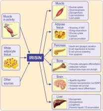
          White adipose tissue (WAT) is an endocrine organ highly integrated in homeostasis and capable of establishing ways of communicating and influencing multiple metabolic processes. Brown adipose tissue promotes energy expenditure by incorporating the uncoupling protein 1 (UCP1), also known as thermogenin, which decouples cellular respiration and heat production, in the mitochondrial membranes. Recent data suggest the presence of a thermogenic cell formation from white adipocytes (beige or brite cells) with a potential role in preventing obesity and metabolic syndrome. The formation of these cells is influenced by physical exertion that induces expression of PPARγ coactivator-1 (PGC1) and downstream membrane protein, fibronectin type III domain-containing protein 5 (FNDC5) in skeletal muscle. Irisin, a thermogenic adipomyokine produced by FNDC5 cleavage is involved in the browning of adipose tissue. While animal studies are congruent with regard to the relationship between physical exertion and irisin release, the results from human studies are less than clear. Therefore, this review focuses on recent advances in our understanding of muscle and adipose tissue thermogenesis. Further, it describes the molecular mechanisms by which irisin impacts exercise, glucose homeostasis and obesity. Finally, the review discusses current gaps and controversies related to irisin release, its mode of action and its future potential as a therapeutic tool in managing obesity and metabolic syndrome.

          Related collections

          Most cited references99

          • Record: found
          • Abstract: found
          • Article: not found

          Adipose tissue, adipokines, and inflammation.

          White adipose tissue is no longer considered an inert tissue mainly devoted to energy storage but is emerging as an active participant in regulating physiologic and pathologic processes, including immunity and inflammation. Macrophages are components of adipose tissue and actively participate in its activities. Furthermore, cross-talk between lymphocytes and adipocytes can lead to immune regulation. Adipose tissue produces and releases a variety of proinflammatory and anti-inflammatory factors, including the adipokines leptin, adiponectin, resistin, and visfatin, as well as cytokines and chemokines, such as TNF-alpha, IL-6, monocyte chemoattractant protein 1, and others. Proinflammatory molecules produced by adipose tissue have been implicated as active participants in the development of insulin resistance and the increased risk of cardiovascular disease associated with obesity. In contrast, reduced leptin levels might predispose to increased susceptibility to infection caused by reduced T-cell responses in malnourished individuals. Altered adipokine levels have been observed in a variety of inflammatory conditions, although their pathogenic role has not been completely clarified.
            Bookmark
            • Record: found
            • Abstract: found
            • Article: not found

            Exercise induces hippocampal BDNF through a PGC-1α/FNDC5 pathway.

            Exercise can improve cognitive function and has been linked to the increased expression of brain-derived neurotrophic factor (BDNF). However, the underlying molecular mechanisms driving the elevation of this neurotrophin remain unknown. Here we show that FNDC5, a previously identified muscle protein that is induced in exercise and is cleaved and secreted as irisin, is also elevated by endurance exercise in the hippocampus of mice. Neuronal Fndc5 gene expression is regulated by PGC-1α, and Pgc1a(-/-) mice show reduced Fndc5 expression in the brain. Forced expression of FNDC5 in primary cortical neurons increases Bdnf expression, whereas RNAi-mediated knockdown of FNDC5 reduces Bdnf. Importantly, peripheral delivery of FNDC5 to the liver via adenoviral vectors, resulting in elevated blood irisin, induces expression of Bdnf and other neuroprotective genes in the hippocampus. Taken together, our findings link endurance exercise and the important metabolic mediators, PGC-1α and FNDC5, with BDNF expression in the brain. Copyright © 2013 Elsevier Inc. All rights reserved.
              Bookmark
              • Record: found
              • Abstract: found
              • Article: not found

              Developmental origin of fat: tracking obesity to its source.

              The development of obesity not only depends on the balance between food intake and caloric utilization but also on the balance between white adipose tissue, which is the primary site of energy storage, and brown adipose tissue, which is specialized for energy expenditure. In addition, some sites of white fat storage in the body are more closely linked than others to the metabolic complications of obesity, such as diabetes. In this Review, we consider how the developmental origins of fat contribute to its physiological, cellular, and molecular heterogeneity and explore how these factors may play a role in the growing epidemic of obesity.
                Bookmark

                Author and article information

                Contributors
                Journal
                Front Endocrinol (Lausanne)
                Front Endocrinol (Lausanne)
                Front. Endocrinol.
                Frontiers in Endocrinology
                Frontiers Media S.A.
                1664-2392
                02 August 2019
                2019
                : 10
                : 524
                Affiliations
                [1] 1Department of Internal Medicine, “Grigore T. Popa” University of Medicine and Pharmacy , Iaşi, Romania
                [2] 2Clinical Hospital “Sf. Spiridon” , Iaşi, Romania
                [3] 3Department of Basic Medical Sciences, College of Osteopathic Medicine, Western University of Health Sciences , Pomona, CA, United States
                [4] 4Department of Health and Human Development, University of Suceava , Suceava, Romania
                Author notes

                Edited by: Reina Villareal, Baylor College of Medicine, United States

                Reviewed by: Claire Joanne Stocker, University of Buckingham, United Kingdom; Alessandra Feraco, San Raffaele Pisana (IRCCS), Italy

                *Correspondence: Mihai Covasa mcovasa@ 123456westernu.edu

                This article was submitted to Obesity, a section of the journal Frontiers in Endocrinology

                Article
                10.3389/fendo.2019.00524
                6687775
                31428053
                c0a6225b-49ea-4d8f-ba6e-c1d50803d24c
                Copyright © 2019 Arhire, Mihalache and Covasa.

                This is an open-access article distributed under the terms of the Creative Commons Attribution License (CC BY). The use, distribution or reproduction in other forums is permitted, provided the original author(s) and the copyright owner(s) are credited and that the original publication in this journal is cited, in accordance with accepted academic practice. No use, distribution or reproduction is permitted which does not comply with these terms.

                History
                : 07 December 2018
                : 16 July 2019
                Page count
                Figures: 2, Tables: 0, Equations: 0, References: 137, Pages: 12, Words: 10032
                Categories
                Endocrinology
                Review

                Endocrinology & Diabetes
                adipose tissue,thermogenesis,adipokines,exercise,skeletal muscle
                Endocrinology & Diabetes
                adipose tissue, thermogenesis, adipokines, exercise, skeletal muscle

                Comments

                Comment on this article