- Record: found
- Abstract: found
- Article: found
Correction: Correction: Reelin controls the positioning of brainstem serotonergic raphe neurons
correction

Read this article at
There is no author summary for this article yet. Authors can add summaries to their articles on ScienceOpen to make them more accessible to a non-specialist audience.
Abstract
There is an error in the Correction published on January 31, 2019. The publisher apologizes
for this error.
The original Fig 7 caption was mistakenly included in the Correction, rather than
the updated Fig 7 caption provided by the author. Please see the complete, correct
Fig 7 caption here:
10.1371/journal.pone.0213449.g001
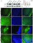
Fig 7
Altered serotonergic innervation of the reeler hippocampus at P20.
(A) Expression of Cxcr4-eGFP (mouse anti-GFP antibody, 1:200, ab38689, Abcam) in Cajal-Retzius
(CR) cells of the WT dentate gyrus (B-D) Serotonergic fibers are distributed throughout
hippocampal layers. (E) Expression of Cxcr4-eGFP in CR cells in reeler hippocampus.
(F-H) Severe reduction of serotonergic fibers in Cxcr4-eGFP hippocampal reeler mice.
CA1, cornu ammonis area 1; DG, dentate gyrus. Scale bar for A-D: 100μm.
Related collections
Most cited references2

- Record: found
- Abstract: found
- Article: found
Reelin controls the positioning of brainstem serotonergic raphe neurons
Reham Shehabeldin, David A. Lutz, Meliha Karsak … (2018)
- Record: found
- Abstract: found
- Article: found
Correction: Reelin controls the positioning of brainstem serotonergic raphe neurons
Reham Shehabeldin, David A. Lutz, Meliha Karsak … (2019)