6
views
0
recommends
+1 Recommend
0 collections
    0
    shares
      • Record: found
      • Abstract: found
      • Article: found
      Is Open Access

      Micro-computed tomography evaluation of the effects of orthodontic force on immature maxillary first molars and alveolar bone mineral density of Sprague–Dawley rats

      research-article

      Read this article at

      Bookmark
          There is no author summary for this article yet. Authors can add summaries to their articles on ScienceOpen to make them more accessible to a non-specialist audience.

          Abstract

          Objective

          To investigate changes in the immature teeth of Sprague–Dawley rats during orthodontic treatment and to explore the changes in the peri-radicular alveolar bone through micro-computed tomography (CT).

          Methods

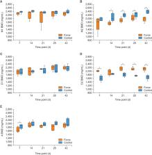
          Twenty-five 26-day-old male Sprague–Dawley rats were included. The maxillary left first molar was moved mesially under a continuous force of 30 cN, and the right first molar served as the control. After orthodontic treatment for 7, 14, 21, 28, and 42 days, the root length, tooth volume, and alveolar bone mineral density (BMD) around the mesial root were measured through micro-CT.

          Results

          The immature teeth continued to elongate after application of orthodontic force. The root length on the force side was significantly smaller than that on the control side, whereas the differences in the volume change between both sides were not statistically significant. Alveolar bone in the coronal part of the compression and tension sides showed no difference in BMD between the experimental and control groups. The BMD of the experimental group decreased from day 14 to day 42 in the apical part of the compression side and increased from day 7 to day 42 in the apical part of the tension side. The BMD of the experimental group decreased in the root apex part on day 7.

          Conclusions

          The root length and volume of immature teeth showed continued development under orthodontic forces. Alveolar bone resorption was observed on the compression side, and bone formation was observed on the tension side.

          Related collections

          Most cited references40

          • Record: found
          • Abstract: found
          • Article: found
          Is Open Access

          State of the Art of Micro-CT Applications in Dental Research

          This review highlights the recent advances in X-ray microcomputed tomography (Micro-CT) applied in dental research. It summarizes Micro-CT applications in measurement of enamel thickness, root canal morphology, evaluation of root canal preparation, craniofacial skeletal structure, micro finite element modeling, dental tissue engineering, mineral density of dental hard tissues and about dental implants. Details of studies in each of these areas are highlighted along with the advantages of Micro-CT, and finally a summary of the future applications of Micro-CT in dental research is given.
            Bookmark
            • Record: found
            • Abstract: found
            • Article: not found

            Evolution of Class III treatment in orthodontics.

            Angle, Tweed, and Moyers classified Class III malocclusions into 3 types: pseudo, dentoalveolar, and skeletal. Clinicians have been trying to identify the best timing to intercept a Class III malocclusion that develops as early as the deciduous dentition. With microimplants as skeletal anchorage, orthopedic growth modification became more effective, and it also increased the scope of camouflage orthodontic treatment for patients who were not eligible for orthognathic surgery. However, orthodontic treatment combined with orthognathic surgery remains the only option for patients with a severe skeletal Class III malocclusion or a craniofacial anomaly. Distraction osteogenesis can now be performed intraorally at an earlier age. The surgery-first approach can minimize the length of time that the malocclusion needs to worsen before orthognathic surgery. Finally, the use of computed tomography scans for 3-dimensional diagnosis and treatment planning together with advances in imaging technology can improve the accuracy of surgical movements and the esthetic outcomes for these patients.
              Bookmark
              • Record: found
              • Abstract: found
              • Article: not found

              Force magnitude and duration effects on amount of tooth movement and root resorption in the rat molar.

              To test the hypothesis that there is no difference in the effect of different continuous moderate to very heavy forces on root resorption or amount of tooth movement. In the study, 10, 25, 50 and 100 g mesial force were applied to the maxillary first molars of rat using nickel titanium closed-coil springs for 3 days, 14 days, and 28 days. The molars were extracted and the surface areas of the root resorption craters were measured using scanning electron microscope. The depths of the root resorption craters were measured using a three-dimensional laser scanning microscope. Tooth movement of the maxillary first molar was measured in relation to the maxillary second molar on digitized lateral cephalometric radiographs. Three days after force application, the tooth movement was not proportionally related to force magnitude. However, 14 days of force application resulted in significantly more tooth movement in the 10, 25, and 50 g force groups than in the 100 g force group. A force application of 10 g produced significantly more tooth movement at 28 days than all the other three force applications. The largest and deepest resorption craters were observed in the disto-buccal root followed by disto-palatal, middle-buccal, middle-palatal, and mesial root. Root resorption and tooth movement increased over time from 3 to 28 days. As heavier forces were applied, greater root resorption occurred. The hypothesis is rejected. The light mesially oriented forces, as applied in this study, produced more tooth movement and less root resorption compared with heavier forces.
                Bookmark

                Author and article information

                Journal
                Korean J Orthod
                Korean J Orthod
                Korean Journal of Orthodontics
                Korean Association of Orthodontists
                2234-7518
                2005-372X
                25 May 2023
                25 May 2023
                25 May 2023
                : 53
                : 3
                : 205-216
                Affiliations
                [1]Department of Orthodontic, Beijing Stomatological Hospital, School of Stomatology, Capital Medical University, Beijing, China
                Author notes
                Corresponding author: Ruofang Zhang. Associate Professor, Department of Orthodontic, Beijing Stomatological Hospital, School of Stomatology, Capital Medical University, Tiantan Xili 4, Dongcheng District, Beijing 100050, China., Tel +86-18610216878 e-mail zhangruofang123@ 123456163.com

                Jingwei Wang and Ruofang Zhang contributed equally to this work (as co-first authors).

                How to cite this article: Wang J, Zhang R, Zhang Z, Geng C, Zhang Y. Micro-computed tomography evaluation of the effects of orthodontic force on immature maxillary first molars and alveolar bone mineral density of Sprague–Dawley rats. Korean J Orthod 2023;53(3):205-216. https://doi.org/10.4041/kjod22.209

                Author information
                https://orcid.org/0000-0001-6508-934X
                https://orcid.org/0000-0001-5849-5729
                Article
                kjod-53-3-205
                10.4041/kjod22.209
                10212771
                37226513
                c6369932-6ea7-4359-b818-f5d2aa7e91a1
                © 2023 The Korean Association of Orthodontists.

                This is an open-access article distributed under the terms of the Creative Commons Attribution Non-Commercial License ( http://creativecommons.org/licenses/by-nc/4.0) which permits unrestricted non-commercial use, distribution, and reproduction in any medium, provided the original work is properly cited.

                History
                : 26 September 2022
                : 16 March 2023
                : 28 April 2023
                Funding
                FUNDING This work was supported by the National Natural Science Foundation of China (Grant No. 81300849), Beijing Natural Science Foundation (Grant No. 7132067), Beijing Municipal Administration of Hospitals Incubating Program (Code: PX2018054), and Scientific Research Foundation for the Returned Overseas Chinese Scholars, Ministry of Education of China (2013).
                Categories
                Original Article

                Dentistry
                root development,alveolar bone,tooth movement
                Dentistry
                root development, alveolar bone, tooth movement

                Comments

                Comment on this article