Inviting an author to review:
Find an author and click ‘Invite to review selected article’ near their name.
Search for authorsSearch for similar articles
5
views
0
recommends
+1 Recommend
0 collections
    0
    shares
      • Record: found
      • Abstract: found
      • Article: found
      Is Open Access

      Clinical application of chromosomal microarray analysis in fetuses with increased nuchal translucency and normal karyotype

      research-article

      Read this article at

      Bookmark
          There is no author summary for this article yet. Authors can add summaries to their articles on ScienceOpen to make them more accessible to a non-specialist audience.

          Abstract

          Background

          Submicroscopic chromosomal imbalance is associated with an increased nuchal translucency (NT). Most previous research has recommended the use of chromosomal microarray analysis (CMA) for prenatal diagnosis if the NT ≥ 3.5 mm. However, there is no current global consensus on the cutoff value for CMA. In this study, we aimed to discuss the fetuses with smaller increased NT which was between cutoff value of NT for karyotype analysis (NT of 2.5 mm in China) and the recommended cutoff value for CMA (NT of 3.5 mm) whether should be excluded from CMA test.

          Methods

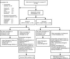
          Singleton pregnant women ( N = 192) who had undergone invasive procedures owing to an increased NT (NT ≥ 2.5 mm) were enrolled. Fetal cells were collected and subjected to single nucleotide polymorphism array and karyotype analyses simultaneously. Cases were excluded if the karyotype analysis indicated aneuploidy and apparent structural aberrations.

          Results

          Fourteen cases of aneuploidy and four cases of structural abnormalities were excluded. Of the remaining 174 cases, 119 fetuses had NTs of 2.5–3.4 mm, and 55 fetuses with NT ≥ 3.5 mm. Eleven copy number variants (CNVs) were identified. In fetuses with smaller NTs, six (6/119, 5.9%) variations were detected, including two (2/119, 1.6%) clinically significant CNVs (pathogenic or likely pathogenic CNV), one  likely benign CNV, two variants unknown significance, and one incidental CNV. Five (5/55, 9.1%) variations were found in fetuses with NT ≥ 3.5 mm. Among these CNVs, three (3/55, 5.5%) cases had clinically significant CNVs, and two had likely benign CNV. There were no statistically significant differences in the incidence of all CNVs and clinically significant CNVs in the two groups ( p > 0.05).

          Conclusion

          CMA improved the diagnostic yield of chromosomal aberrations for fetuses with NTs of 2.5–3.4 mm and apparently normal karyotype, regardless of whether other ultrasonic abnormalities were observed.

          Related collections

          Most cited references22

          • Record: found
          • Abstract: found
          • Article: not found

          Mechanisms of mosaicism, chimerism and uniparental disomy identified by single nucleotide polymorphism array analysis.

          Mosaic aneuploidy and uniparental disomy (UPD) arise from mitotic or meiotic events. There are differences between these mechanisms in terms of (i) impact on embryonic development; (ii) co-occurrence of mosaic trisomy and UPD and (iii) potential recurrence risks. We used a genome-wide single nucleotide polymorphism (SNP) array to study patients with chromosome aneuploidy mosaicism, UPD and one individual with XX/XY chimerism to gain insight into the developmental mechanism and timing of these events. Sixteen cases of mosaic aneuploidy originated mitotically, and these included four rare trisomies and all of the monosomies, consistent with the influence of selective factors. Five trisomies arose meiotically, and three of the five had UPD in the disomic cells, confirming increased risk for UPD in the case of meiotic non-disjunction. Evidence for the meiotic origin of aneuploidy and UPD was seen in the patterns of recombination visible during analysis with 1-3 crossovers per chromosome. The mechanisms of formation of the UPD included trisomy rescue, with and without concomitant trisomy, monosomy rescue, and mitotic formation of a mosaic segmental UPD. UPD was also identified in an XX/XY chimeric individual, with one cell line having complete maternal UPD consistent with a parthenogenetic origin. Utilization of SNP arrays allows simultaneous evaluation of genomic alterations and insights into aneuploidy and UPD mechanisms. Differentiation of mitotic and meiotic origins for aneuploidy and UPD supports existence of selective factors against full trisomy of some chromosomes in the early embryo and provides data for estimation of recurrence and disease mechanisms.
            Bookmark
            • Record: found
            • Abstract: found
            • Article: not found

            Molecular definition of 22q11 deletions in 151 velo-cardio-facial syndrome patients.

            Velo-cardio-facial syndrome (VCFS) is a relatively common developmental disorder characterized by craniofacial anomalies and conotruncal heart defects. Many VCFS patients have hemizygous deletions for a part of 22q11, suggesting that haploinsufficiency in this region is responsible for its etiology. Because most cases of VCFS are sporadic, portions of 22q11 may be prone to rearrangement. To understand the molecular basis for chromosomal deletions, we defined the extent of the deletion, by genotyping 151 VCFS patients and performing haplotype analysis on 105, using 15 consecutive polymorphic markers in 22q11. We found that 83% had a deletion and >90% of these had a similar approximately 3 Mb deletion, suggesting that sequences flanking the common breakpoints are susceptible to rearrangement. We found no correlation between the presence or size of the deletion and the phenotype. To further define the chromosomal breakpoints among the VCFS patients, we developed somatic hybrid cell lines from a set of VCFS patients. An 11-kb resolution physical map of a 1,080-kb region that includes deletion breakpoints was constructed, incorporating genes and expressed sequence tags (ESTs) isolated by the hybridization selection method. The ordered markers were used to examine the two separated copies of chromosome 22 in the somatic hybrid cell lines. In some cases, we were able to map the chromosome breakpoints within a single cosmid. A 480-kb critical region for VCFS has been delineated, including the genes for GSCL, CTP, CLTD, HIRA, and TMVCF, as well as a number of novel ordered ESTs.
              Bookmark
              • Record: found
              • Abstract: found
              • Article: not found

              Increased nuchal translucency with normal karyotype.

              Increased fetal nuchal translucency (NT) thickness between 11 and 14 weeks' gestation is a common phenotypic expression of chromosomal abnormalities, including trisomy 21. However, even in the absence of aneuploidy, nuchal thickening is clinically relevant because it is associated with an increase in adverse perinatal outcome caused by a variety of fetal malformations, dysplasias, deformations, dysruptions, and genetic syndromes. Once the presence of aneuploidy is ruled out, the risk of perinatal outcome dose not statistically increase until the nuchal translucency measurement reaches 3.5 mm or more (>99th percentile). This increase in risk occurs in an exponential fashion as the NT measurement increases. However, if the fetus survives until midgestation, and if a targeted ultrasound at 20 to 22 weeks fails to reveal any abnormalities, the risk of an adverse perinatal outcome and postnatal developmental delay is not statistically increased.
                Bookmark

                Author and article information

                Contributors
                ipxiu304@126.com
                Journal
                Mol Genet Genomic Med
                Mol Genet Genomic Med
                10.1002/(ISSN)2324-9269
                MGG3
                Molecular Genetics & Genomic Medicine
                John Wiley and Sons Inc. (Hoboken )
                2324-9269
                17 June 2019
                August 2019
                : 7
                : 8 ( doiID: 10.1002/mgg3.v7.8 )
                : e811
                Affiliations
                [ 1 ] Fujian Provincial Maternity and Children's Hospital Affiliated Hospital of Fujian Medical University, Fujian Provincial Key Laboratory for Prenatal Diagnosis and Birth Defect Fuzhou China
                Author notes
                [*] [* ] Correspondence

                Liangpu Xu, Fujian Provincial Maternity and Children's Hospital, Affiliated Hospital of Fujian Medical University, Fujian Provincial Key Laboratory for Prenatal Diagnosis and Birth Defect, Fuzhou, China.

                Email: ipxiu304@ 123456126.com

                Author information
                https://orcid.org/0000-0001-7203-0821
                https://orcid.org/0000-0003-2605-2362
                Article
                MGG3811
                10.1002/mgg3.811
                6687862
                31209990
                c65a2af4-7148-4cca-aae8-4926693a0dff
                © 2019 The Authors. Molecular Genetics & Genomic Medicine published by Wiley Periodicals, Inc.

                This is an open access article under the terms of the http://creativecommons.org/licenses/by-nc/4.0/ License, which permits use, distribution and reproduction in any medium, provided the original work is properly cited and is not used for commercial purposes.

                History
                : 20 November 2018
                : 22 May 2019
                : 29 May 2019
                Page count
                Figures: 1, Tables: 3, Pages: 9, Words: 5411
                Funding
                Funded by: The Fujian Provincial Natural Science Foundation
                Award ID: 2017J01238
                Funded by: Research Fund Project of Fujian Provincial Maternity and Children’s Hospital
                Award ID: 2016-25
                Funded by: Fujian Provincial Health and Family Planning Scientific Research Personnel Training Projects
                Award ID: 2017-1-20
                Categories
                Original Article
                Original Articles
                Custom metadata
                2.0
                mgg3811
                August 2019
                Converter:WILEY_ML3GV2_TO_NLMPMC version:5.6.7 mode:remove_FC converted:08.08.2019

                chromosomal microarray analysis,karyotyping,nuchal translucency,prenatal diagnosis

                Comments

                Comment on this article