18
views
0
recommends
+1 Recommend
0 collections
    0
    shares
      • Record: found
      • Abstract: found
      • Article: found
      Is Open Access

      Evaluation of Singh Index and Osteoporosis Self-Assessment Tool for Asians as risk assessment tools of hip fracture in patients with type 2 diabetes mellitus

      research-article

      Read this article at

      Bookmark
          There is no author summary for this article yet. Authors can add summaries to their articles on ScienceOpen to make them more accessible to a non-specialist audience.

          Abstract

          Background

          Type 2 diabetes mellitus (T2DM), an epidemic disease around world, has recently been identified as a risk factor for osteoporosis-associated fracture. However, there is no consensus on the best method of assessing fracture risk in patients with T2DM. The aim of this study was to evaluate the usefulness of the Osteoporosis Self-Assessment Tool for Asians (OSTA) and the Singh Index (SI) in hip fracture risk assessment in patients with T2DM.

          Methods

          We enrolled 261 postmenopausal women with T2DM: 87 had hip fracture resulting from low-energy trauma and 174 age-matched controls had no fracture (two controls per fracture case). Bone mineral density (BMD) was measured with dual-energy X-ray absorptiometry in the lumbar spine and hip region. The SI was obtained from standard antero-posterior radiographs of the pelvis. The OSTA was calculated with a formula based on weight and age. Data were analyzed with descriptive statistics and tests of difference. Receiver operating characteristic analysis was used to determine optimum cutoff values, sensitivity, and specificity of screening methods. Discriminative abilities of different screening tools were compared with the area under the curve (AUC).

          Results

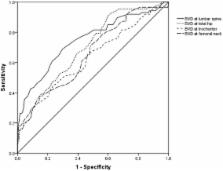
          There were significant differences in BMD at all sites (lumbar spine, femoral neck, trochanter, and total hip) and in SI between the fracture and non-fracture groups ( P < 0.05). There was no significant difference in OSTA between the groups ( P > 0.05). The area under the curve was 0.747 (95% CI: 0.680–0.813) for lumbar spine BMD, 0.699 (95% CI: 0.633–0.764) for total hip BMD, 0.659 (95% CI: 0.589–0.729) for femoral neck BMD, 0.631 (95% CI: 0.557–0.704) for trochanter BMD, 0.534 (95% CI: 0.459–0.610) for OSTA, 0.636 (95% CI: 0.564–0.709) for SI, and 0.795 (95% CI: 0.734–0.857) for OSTA plus SI. The AUC for combined OSTA plus SI was significantly superior to other parameters besides BMD of the lumbar spine.

          Conclusions

          The combination of OSTA plus SI could be a clinical alternative tool for screening of hip fracture risk in large diabetic populations. These tests are inexpensive and simple to perform and could be especially useful in areas where BMD measurement is not accessible.

          Related collections

          Most cited references38

          • Record: found
          • Abstract: found
          • Article: not found

          Association of BMD and FRAX score with risk of fracture in older adults with type 2 diabetes.

          Type 2 diabetes mellitus (DM) is associated with higher bone mineral density (BMD) and paradoxically with increased fracture risk. It is not known if low BMD, central to fracture prediction in older adults, identifies fracture risk in patients with DM. To determine if femoral neck BMD T score and the World Health Organization Fracture Risk Algorithm (FRAX) score are associated with hip and nonspine fracture risk in older adults with type 2 DM. Data from 3 prospective observational studies with adjudicated fracture outcomes (Study of Osteoporotic Fractures [December 1998-July 2008]; Osteoporotic Fractures in Men Study [March 2000-March 2009]; and Health, Aging, and Body Composition study [April 1997-June 2007]) were analyzed in older community-dwelling adults (9449 women and 7436 men) in the United States. Self-reported incident fractures, which were verified by radiology reports. Of 770 women with DM, 84 experienced a hip fracture and 262 a nonspine fracture during a mean (SD) follow-up of 12.6 (5.3) years. Of 1199 men with DM, 32 experienced a hip fracture and 133 a nonspine fracture during a mean (SD) follow-up of 7.5 (2.0) years. Age-adjusted hazard ratios (HRs) for 1-unit decrease in femoral neck BMD T score in women with DM were 1.88 (95% confidence interval [CI], 1.43-2.48) for hip fracture and 1.52 (95% CI, 1.31-1.75) for nonspine fracture, and in men with DM were 5.71 (95% CI, 3.42-9.53) for hip fracture and 2.17 (95% CI, 1.75-2.69) for nonspine fracture. The FRAX score was also associated with fracture risk in participants with DM (HRs for 1-unit increase in FRAX hip fracture score, 1.05; 95% CI, 1.03-1.07, for women with DM and 1.16; 95% CI, 1.07-1.27, for men with DM; HRs for 1-unit increase in FRAX osteoporotic fracture score, 1.04; 95% CI, 1.02-1.05, for women with DM and 1.09; 95% CI, 1.04-1.14, for men with DM). However, for a given T score and age or for a given FRAX score, participants with DM had a higher fracture risk than those without DM. For a similar fracture risk, participants with DM had a higher T score than participants without DM. For hip fracture, the estimated mean difference in T score for women was 0.59 (95% CI, 0.31-0.87) and for men was 0.38 (95% CI, 0.09-0.66). Among older adults with type 2 DM, femoral neck BMD T score and FRAX score were associated with hip and nonspine fracture risk; however, in these patients compared with participants without DM, the fracture risk was higher for a given T score and age or for a given FRAX score.
            Bookmark
            • Record: found
            • Abstract: found
            • Article: not found

            High-resolution peripheral quantitative computed tomographic imaging of cortical and trabecular bone microarchitecture in patients with type 2 diabetes mellitus.

            Cross-sectional epidemiological studies have found that patients with type 2 diabetes mellitus (T2DM) have a higher incidence of certain fragility fractures despite normal or elevated bone mineral density (BMD). In this study, high-resolution peripheral quantitative computed tomography was applied to characterize cortical and trabecular microarchitecture and biomechanics in the peripheral skeleton of female patients with T2DM. A cross-sectional study was conducted in patients with T2DM recruited from a diabetic outpatient clinic. Elderly female patients (age, 62.9 ± 7.7 yr) with a history of T2DM (n = 19) and age- and height-matched controls (n = 19) were recruited. Subjects were imaged using high-resolution peripheral quantitative computed tomography at the distal radius and tibia. Quantitative measures of volumetric (BMD), cross-sectional geometry, trabecular and cortical microarchitecture were calculated. Additionally, compressive mechanical properties were determined by micro-finite element analysis. Compared to the controls, the T2DM cohort had 10% higher trabecular volumetric BMD (P +50%; P < 0.05), whereas pore volume approached significance in the tibia (+118%; P = 0.1). The results of this pilot investigation provide a potential explanation for the inability of standard BMD measures to explain the elevated fracture incidence in patients with T2DM. The findings suggest that T2DM may be associated with impaired resistance to bending loads due to inefficient redistribution of bone mass, characterized by loss of intracortical bone offset by an elevation in trabecular bone density.
              Bookmark
              • Record: found
              • Abstract: found
              • Article: not found

              Association between bone mineral density and type 2 diabetes mellitus: a meta-analysis of observational studies

              Type 2 diabetes mellitus (T2DM) influences bone metabolism, but the relation of T2DM with bone mineral density (BMD) remains inconsistent across studies. The objective of this study was to perform a meta-analysis and meta-regression of the literature to estimate the difference in BMD (g/cm2) between diabetic and non-diabetic populations, and to investigate potential underlying mechanisms. A literature search was performed in PubMed and Ovid extracting data from articles prior to May 2010. Eligible studies were those where the association between T2DM and BMD measured by dual energy X-ray absorptiometry was evaluated using a cross-sectional, cohort or case–control design, including both healthy controls and subjects with T2DM. The analysis was done on 15 observational studies (3,437 diabetics and 19,139 controls). Meta-analysis showed that BMD in diabetics was significantly higher, with pooled mean differences of 0.04 (95% CI: 0.02, 0.05) at the femoral neck, 0.06 (95% CI: 0.04, 0.08) at the hip and 0.06 (95% CI: 0.04, 0.07) at the spine. The differences for forearm BMD were not significantly different between diabetics and non-diabetics. Sex-stratified analyses showed similar results in both genders. Substantial heterogeneity was found to originate from differences in study design and possibly diabetes definition. Also, by applying meta-regression we could establish that younger age, male gender, higher body mass index and higher HbA1C were positively associated with higher BMD levels in diabetic individuals. We conclude that individuals with T2DM from both genders have higher BMD levels, but that multiple factors influence BMD in individuals with T2DM.
                Bookmark

                Author and article information

                Contributors
                bjyyyygkcs@163.com
                doczhyl@163.com
                suineiding@163.com
                fshwxj@163.com
                gukechuangshang@163.com
                (086)01063139264 , doczhyl@hotmail.com
                Journal
                J Orthop Surg Res
                J Orthop Surg Res
                Journal of Orthopaedic Surgery and Research
                BioMed Central (London )
                1749-799X
                3 March 2017
                3 March 2017
                2017
                : 12
                : 37
                Affiliations
                ISNI 0000 0004 0369 153X, GRID grid.24696.3f, Department of Orthopaedics, , Beijing Friendship Hospital, Capital Medical University, ; Yongan Road No.95, Xicheng District, 100050 Beijing, China
                Article
                539
                10.1186/s13018-017-0539-6
                5335822
                28253896
                c6f038b6-825d-4265-939f-5ea1b5414b0a
                © The Author(s). 2017

                Open AccessThis article is distributed under the terms of the Creative Commons Attribution 4.0 International License ( http://creativecommons.org/licenses/by/4.0/), which permits unrestricted use, distribution, and reproduction in any medium, provided you give appropriate credit to the original author(s) and the source, provide a link to the Creative Commons license, and indicate if changes were made. The Creative Commons Public Domain Dedication waiver ( http://creativecommons.org/publicdomain/zero/1.0/) applies to the data made available in this article, unless otherwise stated.

                History
                : 17 November 2016
                : 15 February 2017
                Categories
                Research Article
                Custom metadata
                © The Author(s) 2017

                Surgery
                hip fracture,self-assessment tool for asians,singh index,type 2 diabetes mellitus
                Surgery
                hip fracture, self-assessment tool for asians, singh index, type 2 diabetes mellitus

                Comments

                Comment on this article