59
views
0
recommends
+1 Recommend
0 collections
    0
    shares
      • Record: found
      • Abstract: found
      • Article: found
      Is Open Access

      Expression of miR-21 and its targets (PTEN, PDCD4, TM1) in flat epithelial atypia of the breast in relation to ductal carcinoma in situ and invasive carcinoma

      research-article

      Read this article at

      Bookmark
          There is no author summary for this article yet. Authors can add summaries to their articles on ScienceOpen to make them more accessible to a non-specialist audience.

          Abstract

          Background

          Flat epithelial atypia (FEA) of the breast is characterised by a few layers of mildly atypical luminal epithelial cells. Genetic changes found in ductal carcinoma in situ (DCIS) and invasive ductal breast cancer (IDC) are also found in FEA, albeit at a lower concentration. So far, miRNA expression changes associated with invasive breast cancer, like miR-21, have not been studied in FEA.

          Methods

          We performed miRNA in-situ hybridization (ISH) on 15 cases with simultaneous presence of normal breast tissue, FEA and/or DCIS and 17 additional cases with IDC. Expression of the miR-21 targets PDCD4, TM1 and PTEN was investigated by immunohistochemistry.

          Results

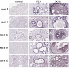
          Two out of fifteen cases showed positive staining for miR-21 in normal breast ductal epithelium, seven out of fifteen cases were positive in the FEA component and nine out of twelve cases were positive in the DCIS component. A positive staining of miR-21 was observed in 15 of 17 IDC cases. In 12 cases all three components were present in one tissue block and an increase of miR-21 from normal breast to FEA and to DCIS was observed in five cases. In three cases the FEA component was negative, whereas the DCIS component was positive for miR-21. In three other cases, normal, FEA and DCIS components were negative for miR-21 and in the last case all three components were positive. Overall we observed a gradual increase in percentage of miR-21 positive cases from normal, to FEA, DCIS and IDC. Immunohistochemical staining for PTEN revealed no obvious changes in staining intensities in normal, FEA, DCIS and IDC. Cytoplasmic staining of PDCD4 increased from normal to IDC, whereas, the nuclear staining decreased. TM1 staining decreased from positive in normal breast to negative in most DCIS and IDC cases. In FEA, the staining pattern for TM1 was similar to normal breast tissue.

          Conclusion

          Upregulation of miR-21 from normal ductal epithelial cells of the breast to FEA, DCIS and IDC parallels morphologically defined carcinogenesis. No clear relation was observed between the staining pattern of miR-21 and its previously reported target genes.

          Related collections

          Most cited references21

          • Record: found
          • Abstract: found
          • Article: not found

          An abundant class of tiny RNAs with probable regulatory roles in Caenorhabditis elegans.

          Two small temporal RNAs (stRNAs), lin-4 and let-7, control developmental timing in Caenorhabditis elegans. We find that these two regulatory RNAs are members of a large class of 21- to 24-nucleotide noncoding RNAs, called microRNAs (miRNAs). We report on 55 previously unknown miRNAs in C. elegans. The miRNAs have diverse expression patterns during development: a let-7 paralog is temporally coexpressed with let-7; miRNAs encoded in a single genomic cluster are coexpressed during embryogenesis; and still other miRNAs are expressed constitutively throughout development. Potential orthologs of several of these miRNA genes were identified in Drosophila and human genomes. The abundance of these tiny RNAs, their expression patterns, and their evolutionary conservation imply that, as a class, miRNAs have broad regulatory functions in animals.
            Bookmark
            • Record: found
            • Abstract: found
            • Article: not found

            MicroRNA-21 is an antiapoptotic factor in human glioblastoma cells.

            MicroRNAs (miRNAs) are small noncoding RNA molecules that regulate protein expression by targeting the mRNA of protein-coding genes for either cleavage or repression of translation. The roles of miRNAs in lineage determination and proliferation as well as the location of several miRNA genes at sites of translocation breakpoints or deletions has led to the speculation that miRNAs could be important factors in the development or maintenance of the neoplastic state. Here we show that the highly malignant human brain tumor, glioblastoma, strongly over-expresses a specific miRNA, miR-21. Our studies show markedly elevated miR-21 levels in human glioblastoma tumor tissues, early-passage glioblastoma cultures, and in six established glioblastoma cell lines (A172, U87, U373, LN229, LN428, and LN308) compared with nonneoplastic fetal and adult brain tissues and compared with cultured nonneoplastic glial cells. Knockdown of miR-21 in cultured glioblastoma cells triggers activation of caspases and leads to increased apoptotic cell death. Our data suggest that aberrantly expressed miR-21 may contribute to the malignant phenotype by blocking expression of critical apoptosis-related genes.
              Bookmark
              • Record: found
              • Abstract: found
              • Article: not found

              MicroRNA-21 targets tumor suppressor genes in invasion and metastasis.

              MicroRNAs (miRNAs) are a class of naturally occurring small non-coding RNAs that target protein-coding mRNAs at the post-transcriptional level. Our previous studies suggest that mir-21 functions as an oncogene and has a role in tumorigenesis, in part through regulation of the tumor suppressor gene tropomyosin 1 (TPM1). Given that TPM1 has been implicated in cell migration, in this study we further investigated the role of mir-21 in cell invasion and tumor metastasis. We found that suppression of mir-21 in metastatic breast cancer MDA-MB-231 cells significantly reduced invasion and lung metastasis. Consistent with this, ectopic expression of TPM1 remarkably reduced cell invasion. Furthermore, we identified two additional direct mir-21 targets, programmed cell death 4 (PDCD4) and maspin, both of which have been implicated in invasion and metastasis. Like TPM1, PDCD4 and maspin also reduced invasiveness of MDA-MB-231 cells. Finally, the expression of PDCD4 and maspin inversely correlated with mir-21 expression in human breast tumor specimens, indicating the potential regulation of PDCD4 and maspin by mir-21 in these tumors. Taken together, the results suggest that, as an oncogenic miRNA, mir-21 has a role not only in tumor growth but also in invasion and tumor metastasis by targeting multiple tumor/metastasis suppressor genes. Therefore, suppression of mir-21 may provide a novel approach for the treatment of advanced cancers.
                Bookmark

                Author and article information

                Journal
                BMC Cancer
                BMC Cancer
                BioMed Central
                1471-2407
                2009
                28 May 2009
                : 9
                : 163
                Affiliations
                [1 ]Department of Pathology & Medical Biology, Groningen University Medical Centre and University of Groningen, Groningen, The Netherlands
                [2 ]Department of Breast Cancer Pathology and Research Laboratory, State Key Laboratory of Breast Cancer Research, Cancer Hospital of Tianjin Medical University, Tianjin, PR China
                Article
                1471-2407-9-163
                10.1186/1471-2407-9-163
                2695476
                19473551
                c782fde9-1a80-4ef2-b12a-76930d2d3572
                Copyright ©2009 Qi et al; licensee BioMed Central Ltd.

                This is an Open Access article distributed under the terms of the Creative Commons Attribution License ( http://creativecommons.org/licenses/by/2.0), which permits unrestricted use, distribution, and reproduction in any medium, provided the original work is properly cited.

                History
                : 12 November 2008
                : 28 May 2009
                Categories
                Research Article

                Oncology & Radiotherapy
                Oncology & Radiotherapy

                Comments

                Comment on this article