0
views
0
recommends
+1 Recommend
0 collections
    0
    shares
      • Record: found
      • Abstract: found
      • Article: found
      Is Open Access

      Different scan areas affect the detection rates of diabetic retinopathy lesions by high-speed ultra-widefield swept-source optical coherence tomography angiography

      research-article

      Read this article at

      Bookmark
          There is no author summary for this article yet. Authors can add summaries to their articles on ScienceOpen to make them more accessible to a non-specialist audience.

          Abstract

          Introduction

          The study aimed to determine the effect of the scanning area used for high-speed ultra-widefield swept-source optical coherence tomography angiography (SS-OCTA) on the detection rate of diabetic retinopathy (DR) lesions.

          Methods

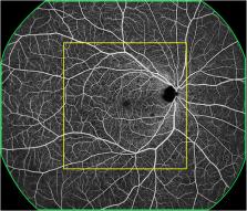
          This prospective, observational study involved diabetic patients between October 2021 and April 2022. The participants underwent a comprehensive ophthalmic examination and high-speed ultra-widefield SS-OCTA using a 24 mm × 20 mm scanning protocol. A central area denoted as “12 mm × 12 mm-central” was extracted from the 24 mm × 20 mm image, and the remaining area was denoted as “12 mm~24mm-annulus.” The rates of detection of DR lesions using the two scanning areas were recorded and compared.

          Results

          In total, 172 eyes (41 eyes with diabetes mellitus without DR, 40 eyes with mild to moderate non-proliferative diabetic retinopathy (NPDR), 51 eyes with severe NPDR, and 40 eyes with proliferative diabetic retinopathy (PDR) from 101 participants were included. The detection rates of microaneurysms (MAs), intraretinal microvascular abnormalities (IRMAs), and neovascularization (NV) for the 12 mm × 12 mm central and 24 mm × 20 mm images were comparable (p > 0.05). The detection rate of NPAs for the 24 mm × 20 mm image was 64.5%, which was significantly higher than that for the 12 mm × 12 mm central image (52.3%, p < 0.05). The average ischemic index (ISI) was 15.26% for the 12 mm~24mm-annulus, which was significantly higher than that for the 12 mm × 12 mm central image (5.62%). Six eyes had NV and 10 eyes had IRMAs that only existed in the 12 mm~24mm-annulus area.

          Conclusions

          The newly developed high-speed ultra-widefield SS-OCTA can capture a 24 mm × 20 mm retinal vascular image during a single scan, which improves the accuracy of detecting the degree of retinal ischemia and detection rate of NV and IRMAs.

          Related collections

          Most cited references35

          • Record: found
          • Abstract: found
          • Article: found
          Is Open Access

          Global Prevalence and Major Risk Factors of Diabetic Retinopathy

          OBJECTIVE To examine the global prevalence and major risk factors for diabetic retinopathy (DR) and vision-threatening diabetic retinopathy (VTDR) among people with diabetes. RESEARCH DESIGN AND METHODS A pooled analysis using individual participant data from population-based studies around the world was performed. A systematic literature review was conducted to identify all population-based studies in general populations or individuals with diabetes who had ascertained DR from retinal photographs. Studies provided data for DR end points, including any DR, proliferative DR, diabetic macular edema, and VTDR, and also major systemic risk factors. Pooled prevalence estimates were directly age-standardized to the 2010 World Diabetes Population aged 20–79 years. RESULTS A total of 35 studies (1980–2008) provided data from 22,896 individuals with diabetes. The overall prevalence was 34.6% (95% CI 34.5–34.8) for any DR, 6.96% (6.87–7.04) for proliferative DR, 6.81% (6.74–6.89) for diabetic macular edema, and 10.2% (10.1–10.3) for VTDR. All DR prevalence end points increased with diabetes duration, hemoglobin A1c, and blood pressure levels and were higher in people with type 1 compared with type 2 diabetes. CONCLUSIONS There are approximately 93 million people with DR, 17 million with proliferative DR, 21 million with diabetic macular edema, and 28 million with VTDR worldwide. Longer diabetes duration and poorer glycemic and blood pressure control are strongly associated with DR. These data highlight the substantial worldwide public health burden of DR and the importance of modifiable risk factors in its occurrence. This study is limited by data pooled from studies at different time points, with different methodologies and population characteristics.
            Bookmark
            • Record: found
            • Abstract: found
            • Article: not found

            Proposed international clinical diabetic retinopathy and diabetic macular edema disease severity scales.

            To develop consensus regarding clinical disease severity classification systems for diabetic retinopathy and diabetic macular edema that can be used around the world, and to improve communication and coordination of care among physicians who care for patients with diabetes. Report regarding the development of clinical diabetic retinopathy disease severity scales. A group of 31 individuals from 16 countries, representing comprehensive ophthalmology, retina subspecialties, endocrinology, and epidemiology. An initial clinical classification system, based on the Early Treatment Diabetic Retinopathy Study and the Wisconsin Epidemiologic Study of Diabetic Retinopathy publications, was circulated to the group in advance of a workshop. Each member reviewed this using e-mail, and a modified Delphi system was used to stratify responses. At a later workshop, separate systems for diabetic retinopathy and macular edema were developed. These were then reevaluated by group members, and the modified Delphi system was again used to measure degrees of agreement. Consensus regarding specific classification systems was achieved. A five-stage disease severity classification for diabetic retinopathy includes three stages of low risk, a fourth stage of severe nonproliferative retinopathy, and a fifth stage of proliferative retinopathy. Diabetic macular edema is classified as apparently present or apparently absent. If training and equipment allow the screener to make a valid decision, macular edema is further categorized as a function of its distance from the central macula. There seems to be a genuine need for consistent international clinical classification systems for diabetic retinopathy and diabetic macular edema that are supported with solid evidence. The proposed clinical classification systems provide a means of appropriately categorizing diabetic retinopathy and macular edema. It is hoped that these systems will be valuable in improving both screening of individuals with diabetes and communication and discussion among individuals caring for these patients.
              Bookmark
              • Record: found
              • Abstract: found
              • Article: not found

              Optical Coherence Tomography Angiography Metrics Predict Progression of Diabetic Retinopathy and Development of Diabetic Macular Edema: A Prospective Study

              To prospectively determine the relationship of OCT angiography (OCTA) metrics to diabetic retinopathy (DR) progression and development of diabetic macular edema (DME).
                Bookmark

                Author and article information

                Contributors
                Journal
                Front Endocrinol (Lausanne)
                Front Endocrinol (Lausanne)
                Front. Endocrinol.
                Frontiers in Endocrinology
                Frontiers Media S.A.
                1664-2392
                20 February 2023
                2023
                : 14
                : 1111360
                Affiliations
                [1] 1 Department of Ophthalmology, Sichuan Provincial People’s Hospital, University of Electronic Science and Technology of China , Chengdu, China
                [2] 2 Department of Ophthalmology, Chinese Academy of Sciences Sichuan Translational Medicine Research Hospital , Chengdu, China
                [3] 3 Eye School, Chengdu University of Traditional Chinese Medicine , Chengdu, China
                [4] 4 School of Medicine, University of Electronic Science and Technology of China , Chengdu, China
                [5] 5 Department of Ophthalmology, Dayi Shaoxiang Hospital , Chengdu, China
                Author notes

                Edited by: Mohd Imtiaz Nawaz, Department of Ophthalmology, King Saud University, Saudi Arabia

                Reviewed by: Salvatore Di Lauro, Hospital Clínico Universitario de Valladolid, Spain; Kai Shi, First Affiliated Hospital of Chongqing Medical University, China; Li Kaiming, The Affiliated Hospital of Southwest Medical University, China

                †These authors have contributed equally to this work and share first authorship

                This article was submitted to Clinical Diabetes, a section of the journal Frontiers in Endocrinology

                Article
                10.3389/fendo.2023.1111360
                9986411
                36891051
                c9889f20-ab47-44ab-80ad-fc889a3d34b7
                Copyright © 2023 Li, Mao, Wei, Liu, Liu, Leng, Wang, Chen, Zhang, Zeng, Wang, Li and Zhong

                This is an open-access article distributed under the terms of the Creative Commons Attribution License (CC BY). The use, distribution or reproduction in other forums is permitted, provided the original author(s) and the copyright owner(s) are credited and that the original publication in this journal is cited, in accordance with accepted academic practice. No use, distribution or reproduction is permitted which does not comply with these terms.

                History
                : 29 November 2022
                : 06 February 2023
                Page count
                Figures: 4, Tables: 4, Equations: 0, References: 35, Pages: 9, Words: 3974
                Funding
                This study was supported in part by the Sichuan Province Science and Technology Support Program (grant number 2021ZYD0108); Health and Family Planning Commission of Sichuan Province (grant number 19ZD012); Science and Technology Project of The Health Planning Committee of Sichuan (grant number 21PJ077); Clinical and Translational Research Fund of Sichuan Provincial People’s Hospital (General Project) (grant number 2020LY04); Sichuan Provincial People’s Hospital (grant number 2021QN13); The Science & Technology Department of Sichuan Province (grant numbers 2017YSKY0001 and 2021YJ0234); and Natural Science Foundation of Sichuan Province (grant number 2023NSFSC0592).
                Categories
                Endocrinology
                Original Research

                Endocrinology & Diabetes
                diabetic retinopathy,microaneurysms,intraretinal microvascular abnormalities,retinal neovascularization,swept-source optical coherence tomography angiography,capillary non-perfusion areas

                Comments

                Comment on this article