9
views
0
recommends
+1 Recommend
0 collections
    0
    shares
      • Record: found
      • Abstract: found
      • Article: found
      Is Open Access

      Patellar maltracking: an update on the diagnosis and treatment strategies

      review-article

      Read this article at

      Bookmark
          There is no author summary for this article yet. Authors can add summaries to their articles on ScienceOpen to make them more accessible to a non-specialist audience.

          Abstract

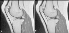
          Patellar maltracking occurs as a result of an imbalance in the dynamic relationship between the patella and trochlea. This is often secondary to an underlying structural abnormality. The clinical evaluation can provide useful clues for the presence of such entity; however, the diagnosis can often be challenging especially in the absence of a documented history of patellar dislocation. Imaging, particularly MRI, can detect subtle features that could lead to the diagnosis, probably even more importantly when there is no clear history of patellar dislocation or before its development. This can provide a road map for formulating a treatment strategy that would be primarily aimed at stabilizing the patellofemoral joint to halt or slow the progression of articular cartilage loss. The purpose of this article is to discuss the clinical and radiologic evaluation of patellar maltracking providing an update on the cross-sectional imaging assessment and also a synopsis of the management options.

          Related collections

          Most cited references71

          • Record: found
          • Abstract: not found
          • Article: not found

          Factors of patellar instability: An anatomic radiographic study

            Bookmark
            • Record: found
            • Abstract: found
            • Article: not found

            Anatomy and biomechanics of the medial patellofemoral ligament.

            The medial patellofemoral ligament (MPFL) is a band of retinacular tissue connecting the femoral medial epicondyle to the medial edge of the patella. The MPFL is approximately 55 mm long, and its width has been reported to range from 3 to 30 mm. The MPFL is overlaid by the distal part of vastus medialis obliquus to a variable extent, and fibres of MPFL merge into the deep aspect of the muscle. Despite the MPFL being very thin, it had a mean tensile strength of 208 N, and has been reported to be the primary passive restraint to patellar lateral displacement. Lateral patellar displacement tests in vitro showed that the patella subluxed most easily at 20 degrees knee flexion. The contribution of the MPFL to resisting patellar lateral subluxation was greatest in the extended knee. This finding was linked to the retinaculae being tightest in full knee extension, and slackening with flexion.
              Bookmark
              • Record: found
              • Abstract: found
              • Article: not found

              Acute lateral patellar dislocation at MR imaging: injury patterns of medial patellar soft-tissue restraints and osteochondral injuries of the inferomedial patella.

              To assess magnetic resonance (MR) imaging findings after acute lateral patellar dislocation (LPD) with emphasis on the medial patella restraints and to describe a medial patellar impaction deformity. Knee MR images obtained within 8 weeks after LPD were evaluated for medial retinacular and medial patellofemoral ligament (MPFL) disruption, vastus medialis obliquus (VMO) edema and/or elevation, and other derangements. One hundred patients with no evidence of prior LPD were evaluated as controls. The Student t test was used for statistical comparisons. Eighty-two examinations were performed in 81 patients with LPD (mean age, 20 years; age range, 9-57 years). Seventy-six percent (62 of 82 examinations) showed medial retinacular disruption at its patellar insertion; 30% (25 of 82), at its midsubstance. The MPFL femoral origin was identified in 87% (71 of 82); of these, 49% (35 of 71) showed injury. Forty-eight percent (39 of 82) showed more than one site of injury to the medial stabilizers; 45% (37 of 82) showed edema or hemorrhage at the inferior VMO. Mean VMO elevation in the coronal plane of the adductor tendon was 2.2 cm, with a range of 0.6-4.5 cm (in control subjects, 0.9 cm; range, 0.1-2.5 cm; P <.001). At the inferomedial patella, 70% (57 of 82) of LPD examinations showed osteochondral injury and 44% (36 of 82) showed concave impaction deformity (0 of 100 control subjects). Other examination findings in LPDs included contusions of the lateral femoral condyle (66 [80%] of 82 examinations) or medial patella (50 [61%] of 82), intraarticular bodies (12 [15%] of 82), effusion (45 [55%] of 82), medial collateral injury (nine [11%] of 82), and meniscal tear (nine [11%] of 82). Injury to the medial retinaculum, MPFL, and VMO may be identified at MR imaging after acute LPD. Concave impaction deformity of the inferomedial patella is a specific sign of prior LPD.
                Bookmark

                Author and article information

                Contributors
                613-737-8899 , zjibri@toh.ca
                Journal
                Insights Imaging
                Insights Imaging
                Insights into Imaging
                Springer Berlin Heidelberg (Berlin/Heidelberg )
                1869-4101
                14 June 2019
                14 June 2019
                December 2019
                : 10
                : 65
                Affiliations
                [1 ]ISNI 0000 0000 9606 5108, GRID grid.412687.e, Department of Medical Imaging, , The Ottawa Hospital, ; 501 Smyth Road, Ottawa, ON K1H 8L6 Canada
                [2 ]ISNI 0000 0001 2182 2255, GRID grid.28046.38, Faculty of Medicine, , University of Ottawa, ; 451 Smyth Road, Ottawa, ON K1H 8M5 Canada
                [3 ]ISNI 0000 0000 9606 5108, GRID grid.412687.e, Division of Orthopaedic Surgery, , The Ottawa Hospital, ; 501 Smyth Road, Ottawa, ON K1H 8L6 Canada
                Article
                755
                10.1186/s13244-019-0755-1
                6570735
                31201575
                ca4d765b-f419-4fc1-a6b9-4c059d0b19fa
                © The Author(s). 2019

                Open AccessThis article is distributed under the terms of the Creative Commons Attribution 4.0 International License ( http://creativecommons.org/licenses/by/4.0/), which permits unrestricted use, distribution, and reproduction in any medium, provided you give appropriate credit to the original author(s) and the source, provide a link to the Creative Commons license, and indicate if changes were made.

                History
                : 24 January 2019
                : 21 May 2019
                Categories
                Educational Review
                Custom metadata
                © The Author(s) 2019

                Radiology & Imaging
                patellar maltracking,patellar instability,anterior knee pain,patellofemoral osteoarthritis

                Comments

                Comment on this article