16
views
0
recommends
+1 Recommend
0 collections
    0
    shares
      • Record: found
      • Abstract: found
      • Article: found
      Is Open Access

      Modulation of cue-induced firing of ventral tegmental area dopamine neurons by leptin and ghrelin

      research-article

      Read this article at

      Bookmark
          There is no author summary for this article yet. Authors can add summaries to their articles on ScienceOpen to make them more accessible to a non-specialist audience.

          Abstract

          Background/objectives:

          The rewarding value of palatable foods contributes to overconsumption, even in satiated subjects. Midbrain dopaminergic activity in response to reward-predicting environmental stimuli drives reward-seeking and motivated behavior for food rewards. This mesolimbic dopamine (DA) system is sensitive to changes in energy balance, yet it has thus far not been established whether reward signaling of DA neurons in vivo is under control of hormones that signal appetite and energy balance such as ghrelin and leptin.

          Subjects/methods:

          We trained rats ( n=11) on an operant task in which they could earn two different food rewards. We then implanted recording electrodes in the ventral tegmental area (VTA), and recorded from DA neurons during behavior. Subsequently, we assessed the effects of mild food restriction and pretreatment with the adipose tissue-derived anorexigenic hormone leptin or the orexigenic hormone ghrelin on VTA DA reward signaling.

          Results:

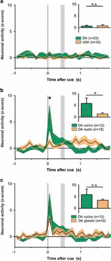
          Animals showed an increase in performance following mild food restriction ( P=0.002). Importantly, food-cue induced DA firing increased when animals were food restricted ( P=0.02), but was significantly attenuated after leptin pretreatment ( P=0.00). While ghrelin did affect baseline DA activity ( P=0.025), it did not affect cue-induced firing ( P⩾0.353).

          Conclusions:

          Metabolic signals, such as leptin, affect food seeking, a process that is dependent on the formation of cue-reward outcomes and involves midbrain DA signaling. These data show that food restriction engages the encoding of food cues by VTA DA neurons at a millisecond level and leptin suppresses this activity. This suggests that leptin is a key in linking metabolic information to reward signaling.

          Related collections

          Most cited references36

          • Record: found
          • Abstract: found
          • Article: not found

          Leptin receptor signaling in midbrain dopamine neurons regulates feeding.

          The leptin hormone is critical for normal food intake and metabolism. While leptin receptor (Lepr) function has been well studied in the hypothalamus, the functional relevance of Lepr expression in the ventral tegmental area (VTA) has not been investigated. The VTA contains dopamine neurons that are important in modulating motivated behavior, addiction, and reward. Here, we show that VTA dopamine neurons express Lepr mRNA and respond to leptin with activation of an intracellular JAK-STAT pathway and a reduction in firing rate. Direct administration of leptin to the VTA caused decreased food intake while long-term RNAi-mediated knockdown of Lepr in the VTA led to increased food intake, locomotor activity, and sensitivity to highly palatable food. These data support a critical role for VTA Lepr in regulating feeding behavior and provide functional evidence for direct action of a peripheral metabolic signal on VTA dopamine neurons.
            Bookmark
            • Record: found
            • Abstract: found
            • Article: not found

            The control of firing pattern in nigral dopamine neurons: burst firing.

            In addition to firing in a single spiking mode, dopamine (DA) cells have been observed to fire in a bursting pattern with consecutive spikes in a burst displaying progressively decreasing amplitude and increasing duration. In vivo intracellular recording demonstrated the bursts to typically ride on a depolarizing wave (5 to 15 mV amplitude). Although the burst-firing frequency of DA cells showed little correlation with the base line firing rate, increases in firing rate were usually associated with an increase in burst firing. Increases in burst firing could also be elicited by intracellular calcium injection and could be prevented by intracellular injection of EGTA, suggesting a calcium involvement in bursting. Blockade of potassium conductances with extracellular iontophoresis of barium or intracellular injection of tetraethylammonium bromide could also trigger an increased degree of burst firing in DA cells. These data suggest that the increased calcium influx accompanying an increased firing rate triggers burst firing, possibly by inactivating a potassium conductance. A switch from a single spiking mode to a burst-firing mode may be important in modulating striatal DA release, as shown for burst firing in other preparations.
              Bookmark
              • Record: found
              • Abstract: found
              • Article: not found

              Responses of monkey dopamine neurons to reward and conditioned stimuli during successive steps of learning a delayed response task.

              The present investigation had two aims: (1) to study responses of dopamine neurons to stimuli with attentional and motivational significance during several steps of learning a behavioral task, and (2) to study the activity of dopamine neurons during the performance of cognitive tasks known to be impaired after lesions of these neurons. Monkeys that had previously learned a simple reaction time task were trained to perform a spatial delayed response task via two intermediate tasks. During the learning of each new task, a total of 25% of 76 dopamine neurons showed phasic responses to the delivery of primary liquid reward, whereas only 9% of 163 neurons responded to this event once task performance was established. This produced an average population response during but not after learning of each task. Reward responses during learning were significantly more numerous and pronounced in area A10, as compared to areas A8 and A9. Dopamine neurons also showed phasic responses to the two conditioned stimuli. These were the instruction cue, which was the first stimulus in each trial and indicated the target of the upcoming arm movement (58% of 76 neurons during and 44% of 163 neurons after learning), and the trigger stimulus, which was a conditioned incentive stimulus predicting reward and eliciting a saccadic eye movement and an arm reaching movement (38% of neurons during and 40% after learning). None of the dopamine neurons showed sustained activity in the delay between the instruction and trigger stimuli that would resemble the activity of neurons in dopamine terminal areas, such as the striatum and frontal cortex. Thus, dopamine neurons respond phasically to alerting external stimuli with behavioral significance whose detection is crucial for learning and performing delayed response tasks. The lack of sustained activity suggests that dopamine neurons do not encode representational processes, such as working memory, expectation of external stimuli or reward, or preparation of movement. Rather, dopamine neurons are involved with transient changes of impulse activity in basic attentional and motivational processes underlying learning and cognitive behavior.
                Bookmark

                Author and article information

                Journal
                Int J Obes (Lond)
                Int J Obes (Lond)
                International Journal of Obesity (2005)
                Nature Publishing Group
                0307-0565
                1476-5497
                December 2015
                17 July 2015
                11 August 2015
                : 39
                : 12
                : 1742-1749
                Affiliations
                [1 ]Department of Translational Neuroscience, Brain Center Rudolf Magnus, University Medical Center Utrecht , Utrecht, The Netherlands
                [2 ]Master's Programme Neuroscience and Cognition, Utrecht University , Utrecht, The Netherlands
                [3 ]Department of Psychiatry & Cell Biology and Physiology, UNC Neuroscience Center, University of North Carolina at Chapel Hill , Chapel Hill, NC, USA
                Author notes
                [* ]Department of Translational Neuroscience, Brain Center Rudolf Magnus , Universiteitsweg 100, Utrecht 3584 CG, The Netherlands. E-mail: g.vanderplasse@ 123456umcutrecht.nl
                [4]

                These authors contributed equally to this work.

                Article
                ijo2015131
                10.1038/ijo.2015.131
                4722241
                26183405
                cab0552f-304d-432e-ac98-72e9ccdc334e
                Copyright © 2015 Macmillan Publishers Limited

                This work is licensed under a Creative Commons Attribution 4.0 International License. The images or other third party material in this article are included in the article's Creative Commons license, unless indicated otherwise in the credit line; if the material is not included under the Creative Commons license, users will need to obtain permission from the license holder to reproduce the material. To view a copy of this license, visit http://creativecommons.org/licenses/by/4.0/

                History
                : 18 February 2015
                : 08 June 2015
                : 12 July 2015
                Categories
                Original Article

                Nutrition & Dietetics
                Nutrition & Dietetics

                Comments

                Comment on this article