Inviting an author to review:
Find an author and click ‘Invite to review selected article’ near their name.
Search for authorsSearch for similar articles
0
views
0
recommends
+1 Recommend
0 collections
    0
    shares
      • Record: found
      • Abstract: found
      • Article: found
      Is Open Access

      A New Technique for the Endoscopic Reconstruction of Skull Base Defects Using Multiple-balloon Catheters

      methods-article

      Read this article at

      Bookmark
          There is no author summary for this article yet. Authors can add summaries to their articles on ScienceOpen to make them more accessible to a non-specialist audience.

          Abstract

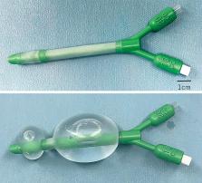
          Cerebrospinal fluid (CSF) leakage is a major complication following endoscopic endonasal skull base surgery. Various skull base reconstruction methods are available, and the use of a vascularized nasoseptal flap (NSF) in skull base reconstruction has greatly contributed to a decrease in the CSF leak rate. A balloon catheter such as a sinus balloon or a Foley catheter is often used to support an NSF; however, in cases wherein nasal and/or paranasal structures supporting the balloon are lacking following the surgery, the NSF is not properly fixed and postoperative CSF leak may occur. Here we introduce a new technique of using multiple-balloon catheters to fix an NSF in such cases and provide the results of our analysis of the new technique's efficacy. Eight patients who underwent endonasal endoscopic surgery for the following cases were included: olfactory neuroblastoma (n = 6), recurrent craniofacial meningioma (n = 1), and recurrent chordoma (n = 1). After tumor resection, multilayered reconstruction with vascularized NSF was performed. Given that the Foley catheter was not stable to fix the flap in each case, we used an additional nasal catheter to support the Foley catheter. No complications such as postoperative CSF leak and necrosis of the vascularized flap were observed. These results suggest that the multiple-balloon catheter technique is a useful method for fixing the NSF to the skull base even when nasal cavity structures are missing due to surgical removal.

          Related collections

          Most cited references13

          • Record: found
          • Abstract: found
          • Article: not found

          A novel reconstructive technique after endoscopic expanded endonasal approaches: vascular pedicle nasoseptal flap.

          In patients with large dural defects of the anterior and ventral skull base after endonasal skull base surgery, there is a significant risk of a postoperative cerebrospinal fluid leak after reconstruction. Reconstruction with vascularized tissue is desirable to facilitate rapid healing, especially in irradiated patients. We developed a neurovascular pedicled flap of the nasal septum mucoperiosteum and mucoperichondrium based on the nasoseptal artery, a branch of the posterior septal artery (Hadad-Bassagasteguy flap [HBF]). A retrospective review of patients undergoing endonasal skull base surgery at the University of Rosario, Argentina, and the University of Pittsburgh Medical Center was performed to identify patients who were reconstructed with a vascularized septal mucosal flap. Forty-three patients undergoing endonasal cranial base surgery were repaired with the septal mucosal flap. Two patients with postoperative cerebrospinal fluid leaks (5%) were successfully treated with focal fat grafts. We encountered no infectious or wound complications in this series of patients. One patient experienced a posterior nose bleed from the posterior nasal artery. This was controlled with electrocautery and the flap blood supply was preserved. The HBF is a versatile and reliable reconstructive technique for defects of the anterior, middle, clival, and parasellar skull base. Its use has resulted in a sharp decrease in the incidence of postoperative cerebrospinal fluid leaks after endonasal skull base surgery and is recommended for the reconstruction of large dural defects and when postoperative radiation therapy is anticipated.
            Bookmark
            • Record: found
            • Abstract: found
            • Article: not found

            Expanded endonasal approach: fully endoscopic, completely transnasal approach to the middle third of the clivus, petrous bone, middle cranial fossa, and infratemporal fossa.

            The middle third of the clivus and the region around the petrous internal carotid artery (ICA) is a difficult area of the skull base in terms of access. This is a deep area rich with critical neurovascular structures, which is often host to typical skull base diseases. Expanded endoscopic endonasal approaches offer a potential option for accessing this difficult region. The objective of this paper was to establish the clinical feasibility of gaining access to the paraclival space in the region of the middle third of the clivus, to provide a practical modular and clinically applicable classification, and to describe the relevant critical surgical anatomy for each module. The anatomical organization of the region around the petrous ICA, cavernous sinus, and middle clivus is presented, with approaches divided into zones. In an accompanying paper in this issue by Cavallo, et al., the anatomy of the pterygopalatine fossa is presented; this was observed through cadaveric dissection for which an expanded endonasal approach was used. In the current paper the authors translate the aforementioned anatomical study to provide a clinically applicable categorization of the endonasal approach to the region around the petrous ICA. A series of zones inferior and superior to the petrous ICA are described, with an illustrative case presented for each region. The expanded endonasal approach is a feasible approach to the middle third of the clivus, petrous ICA, cavernous sinus, and medial infratemporal fossa in cases in which the lesion is located centrally, with neurovascular structures displaced laterally.
              Bookmark
              • Record: found
              • Abstract: found
              • Article: not found

              Endoscopic endonasal skull base reconstruction using a nasal septal flap: surgical results and comparison with previous reconstructions.

              The objective of this study is to evaluate the usefulness and reliability of endoscopic endonasal skull base reconstructions using a nasal septal flap. This study is designed as a retrospective review. Between April 2005 and November 2009, we performed 32 endoscopic endonasal skull base reconstructions for closure of large dural defects. Eleven patients underwent reconstructions using fat grafts or the fascia lata (non-flap group). Twenty one patients underwent reconstructions using a nasal septal flap with a balloon catheter (flap group). Incidence of postoperative cerebrospinal fluid (CSF) leaks and perioperative insertion rate of external lumbar drain (ELD) were compared between the two groups. Postoperative CSF leaks occurred in two patients (9.5%) in the flap group. Three patients (27.3%) presented CSF leaks in the non-flap group. The rate of insertion of ELD was 81.8% in the non-flap group. In the flap group, one patient (4.8%) should be placed with ELD postoperatively. The incidence of postoperative CSF leaks in the flap group was lower than in the non-flap group, whereas the rate of insertion of ELD in the non-flap group was higher than in the flap group. Endoscopic endonasal skull base reconstruction using a nasal septal flap without ELD seems to be useful and reliable for ventral skull base defects after endoscopic endonasal approaches as compared with our previous single-layer reconstructions using free fat grafts or fascia lata. The long-term effectiveness of nasal septal flaps to prevent intracranial complications should be confirmed.
                Bookmark

                Author and article information

                Journal
                Neurol Med Chir (Tokyo)
                Neurol Med Chir (Tokyo)
                Neurologia medico-chirurgica
                The Japan Neurosurgical Society
                0470-8105
                1349-8029
                6 September 2022
                October 2022
                : 62
                : 10
                : 483-487
                Affiliations
                [1 ]Department of Neurosurgery, Tokyo Medical and Dental University, Tokyo, Japan
                [2 ]Department of Head and Neck Surgery, Tokyo Medical and Dental University, Tokyo, Japan
                Author notes

                Corresponding author: Yoji Tanaka, MD, Ph.D

                Department of Neurosurgery, Tokyo Medical and Dental University, 1-5-45 Yushima, Bunkyo-ku, Tokyo 113-8510, Japan.

                e-mail: tanaka.nsrg@ 123456tmd.ac.jp

                Article
                10.2176/jns-nmc.2022-0146
                9637399
                36070960
                cbeadc04-72fd-49df-8cfd-1ab1516d42b6
                © 2022 The Japan Neurosurgical Society

                This work is licensed under a Creative Commons Attribution-NonCommercial-NoDerivatives International License.

                History
                : 13 May 2022
                : 24 June 2022
                Categories
                Technical Note

                neuroendoscopy,skull base reconstruction,balloon catheter,cerebrospinal fluid leak

                Comments

                Comment on this article