4
views
0
recommends
+1 Recommend
0 collections
    0
    shares
      • Record: found
      • Abstract: found
      • Article: found
      Is Open Access

      Application of the 3D slicer chest imaging platform segmentation algorithm for large lung nodule delineation

      research-article

      Read this article at

      Bookmark
          There is no author summary for this article yet. Authors can add summaries to their articles on ScienceOpen to make them more accessible to a non-specialist audience.

          Abstract

          Purpose

          Accurate segmentation of lung nodules is crucial in the development of imaging biomarkers for predicting malignancy of the nodules. Manual segmentation is time consuming and affected by inter-observer variability. We evaluated the robustness and accuracy of a publically available semiautomatic segmentation algorithm that is implemented in the 3D Slicer Chest Imaging Platform (CIP) and compared it with the performance of manual segmentation.

          Methods

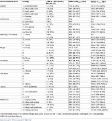
          CT images of 354 manually segmented nodules were downloaded from the LIDC database. Four radiologists performed the manual segmentation and assessed various nodule characteristics. The semiautomatic CIP segmentation was initialized using the centroid of the manual segmentations, thereby generating four contours for each nodule. The robustness of both segmentation methods was assessed using the region of uncertainty (δ) and Dice similarity index (DSI). The robustness of the segmentation methods was compared using the Wilcoxon-signed rank test (p Wilcoxon<0.05). The Dice similarity index (DSI Agree) between the manual and CIP segmentations was computed to estimate the accuracy of the semiautomatic contours.

          Results

          The median computational time of the CIP segmentation was 10 s. The median CIP and manually segmented volumes were 477 ml and 309 ml, respectively. CIP segmentations were significantly more robust than manual segmentations (median δ CIP = 14ml, median dsi CIP = 99% vs. median δ manual = 222ml, median dsi manual = 82%) with p Wilcoxon~10 −16. The agreement between CIP and manual segmentations had a median DSI Agree of 60%. While 13% (47/354) of the nodules did not require any manual adjustment, minor to substantial manual adjustments were needed for 87% (305/354) of the nodules. CIP segmentations were observed to perform poorly (median DSI Agree≈50%) for non-/sub-solid nodules with subtle appearances and poorly defined boundaries.

          Conclusion

          Semi-automatic CIP segmentation can potentially reduce the physician workload for 13% of nodules owing to its computational efficiency and superior stability compared to manual segmentation. Although manual adjustment is needed for many cases, CIP segmentation provides a preliminary contour for physicians as a starting point.

          Related collections

          Most cited references36

          • Record: found
          • Abstract: found
          • Article: not found

          The National Lung Screening Trial: overview and study design.

          The National Lung Screening Trial (NLST) is a randomized multicenter study comparing low-dose helical computed tomography (CT) with chest radiography in the screening of older current and former heavy smokers for early detection of lung cancer, which is the leading cause of cancer-related death in the United States. Five-year survival rates approach 70% with surgical resection of stage IA disease; however, more than 75% of individuals have incurable locally advanced or metastatic disease, the latter having a 5-year survival of less than 5%. It is plausible that treatment should be more effective and the likelihood of death decreased if asymptomatic lung cancer is detected through screening early enough in its preclinical phase. For these reasons, there is intense interest and intuitive appeal in lung cancer screening with low-dose CT. The use of survival as the determinant of screening effectiveness is, however, confounded by the well-described biases of lead time, length, and overdiagnosis. Despite previous attempts, no test has been shown to reduce lung cancer mortality, an endpoint that circumvents screening biases and provides a definitive measure of benefit when assessed in a randomized controlled trial that enables comparison of mortality rates between screened individuals and a control group that does not undergo the screening intervention of interest. The NLST is such a trial. The rationale for and design of the NLST are presented. © RSNA, 2010
            Bookmark
            • Record: found
            • Abstract: found
            • Article: not found

            Three-dimensional multi-scale line filter for segmentation and visualization of curvilinear structures in medical images.

            This paper describes a method for the enhancement of curvilinear structures such as vessels and bronchi in three-dimensional (3-D) medical images. A 3-D line enhancement filter is developed with the aim of discriminating line structures from other structures and recovering line structures of various widths. The 3-D line filter is based on a combination of the eigenvalues of the 3-D Hessian matrix. Multi-scale integration is formulated by taking the maximum among single-scale filter responses, and its characteristics are examined to derive criteria for the selection of parameters in the formulation. The resultant multi-scale line-filtered images provide significantly improved segmentation and visualization of curvilinear structures. The usefulness of the method is demonstrated by the segmentation and visualization of brain vessels from magnetic resonance imaging (MRI) and magnetic resonance angiography (MRA), bronchi from a chest CT, and liver vessels (portal veins) from an abdominal CT.
              Bookmark
              • Record: found
              • Abstract: found
              • Article: not found

              Solitary pulmonary nodules: Part I. Morphologic evaluation for differentiation of benign and malignant lesions.

              The solitary pulmonary nodule is a common radiologic abnormality that is often detected incidentally. Although most solitary pulmonary nodules have benign causes, many represent stage I lung cancers and must be distinguished from benign nodules in an expeditious and cost-effective manner. Evaluation of specific morphologic features of a solitary pulmonary nodule with conventional imaging techniques can help differentiate benign from malignant nodules and obviate further costly assessment. Small size and smooth, well-defined margins are suggestive of but not diagnostic for benignity. Lobulated contour as well as an irregular or spiculated margin with distortion of adjacent vessels are typically associated with malignancy. There is considerable overlap in the internal characteristics (eg, attenuation, cavitation, wall thickness) of benign and malignant nodules. The presence of intranodular fat is a reliable indicator of a hamartoma. The presence and pattern of calcification can also help differentiate benign from malignant nodules. Computed tomography (CT) (particularly thin-section CT) is 10-20 times more sensitive than standard radiography and allows objective, quantitative assessment of calcification. Initial evaluation often results in nonspecific findings, in which case nodules are classified as indeterminate and require further evaluation to exclude malignancy. Growth rate assessment, Bayesian analysis, contrast material-enhanced CT, positron emission tomography, and transthoracic needle aspiration biopsy can be useful in this regard.
                Bookmark

                Author and article information

                Contributors
                Role: Editor
                Journal
                PLoS One
                PLoS ONE
                plos
                plosone
                PLoS ONE
                Public Library of Science (San Francisco, CA USA )
                1932-6203
                8 June 2017
                2017
                : 12
                : 6
                : e0178944
                Affiliations
                [1 ]Department of Radiation Oncology, Dana-Farber Cancer Institute, Brigham and Women’s Hospital, and Harvard Medical School, Boston, MA, United States of America
                [2 ]Biomedical Engineering Department, Mayo Graduate School of Medicine Rochester, MN, United States of America
                [3 ]Department of Radiology, Brigham and Women’s Hospital and Harvard Medical School, Boston, MA, United States of America
                [4 ]Isomics, Inc., Cambridge, MA, United States of America
                [5 ]Department of Radiology, University of Michigan Health System, Ann Arbor MI, United States of America
                University of Groningen, University Medical Center Groningen, NETHERLANDS
                Author notes

                Competing Interests: Dr. Steve Pieper is the owner and an employee of Isomics, Inc., but Isomics, Inc. is not a funder of this study. There are no patents, products in development or marketed products to declare. This does not alter our adherence to all the PLOS ONE policies on sharing data and materials, as detailed online in the guide for authors.

                • Conceptualization: SSFY CP HJWLA.

                • Data curation: DB SP SSFY CP HJWLA.

                • Formal analysis: SSFY CP JK HJWLA.

                • Funding acquisition: HJWLA RSJE.

                • Investigation: SSFY CP DB JK.

                • Methodology: SSFY CP RSJE SP JK HJWLA.

                • Project administration: SSFY CP DB SP HJWLA.

                • Resources: DB RSJE SP.

                • Software: DB RSJE SP.

                • Supervision: DB SP HJWLA.

                • Validation: SSFY CP JK HJWLA.

                • Visualization: SSFY CP JK.

                • Writing – original draft: SSFY CP HJWLA.

                • Writing – review & editing: SSFY CP DB RSJE SP JK HJWLA.

                Author information
                http://orcid.org/0000-0001-8626-5736
                Article
                PONE-D-17-05855
                10.1371/journal.pone.0178944
                5464594
                28594880
                cdf04162-ed47-49d6-95cc-87fba535e533
                © 2017 Yip et al

                This is an open access article distributed under the terms of the Creative Commons Attribution License, which permits unrestricted use, distribution, and reproduction in any medium, provided the original author and source are credited.

                History
                : 13 February 2017
                : 22 May 2017
                Page count
                Figures: 5, Tables: 3, Pages: 17
                Funding
                Funded by: funder-id http://dx.doi.org/10.13039/100000054, National Cancer Institute;
                Award ID: U01CA190234
                Award Recipient :
                Funded by: funder-id http://dx.doi.org/10.13039/100000054, National Cancer Institute;
                Award ID: U24CA194354
                Award Recipient :
                Funded by: funder-id http://dx.doi.org/10.13039/100000002, National Institutes of Health;
                Award ID: 1R01HL116931
                Award Recipient :
                The authors would like to acknowledge support from the National Institutes of Health (award number U01CA190234 and U24CA194354) and research seed funding grant from the American Association of Physicists in Medicine. The Chest Imaging Platform (CIP) is supported by the National Institutes of Health award number 1R01HL116931. Isomics, Inc. provided support in the form of salaries for author SP, but did not have any additional role in the study design, data collection and analysis, decision to publish, or preparation of the manuscript. The specific role of this author is articulated in the ‘author contributions’ section.
                Categories
                Research Article
                People and Places
                Population Groupings
                Professions
                Radiologists
                Medicine and Health Sciences
                Diagnostic Medicine
                Diagnostic Radiology
                Pulmonary Imaging
                Research and Analysis Methods
                Imaging Techniques
                Diagnostic Radiology
                Pulmonary Imaging
                Medicine and Health Sciences
                Radiology and Imaging
                Diagnostic Radiology
                Pulmonary Imaging
                Physical Sciences
                Mathematics
                Applied Mathematics
                Algorithms
                Research and Analysis Methods
                Simulation and Modeling
                Algorithms
                Medicine and Health Sciences
                Oncology
                Cancers and Neoplasms
                Lung and Intrathoracic Tumors
                Biology and Life Sciences
                Developmental Biology
                Morphogenesis
                Morphogenic Segmentation
                Biology and Life Sciences
                Physiology
                Physiological Processes
                Calcification
                Medicine and Health Sciences
                Physiology
                Physiological Processes
                Calcification
                Research and Analysis Methods
                Imaging Techniques
                Neuroimaging
                Computed Axial Tomography
                Biology and Life Sciences
                Neuroscience
                Neuroimaging
                Computed Axial Tomography
                Medicine and Health Sciences
                Diagnostic Medicine
                Diagnostic Radiology
                Tomography
                Computed Axial Tomography
                Research and Analysis Methods
                Imaging Techniques
                Diagnostic Radiology
                Tomography
                Computed Axial Tomography
                Medicine and Health Sciences
                Radiology and Imaging
                Diagnostic Radiology
                Tomography
                Computed Axial Tomography
                Medicine and Health Sciences
                Health Care
                Health Care Providers
                Medical Doctors
                Physicians
                People and Places
                Population Groupings
                Professions
                Medical Doctors
                Physicians
                Custom metadata
                Since a publicly available dataset was used in this study, approval by an institutional review board was not needed. A publicly available thoracic CT dataset, known as the Lung Image Database Consortium (LIDC), was downloaded from The Cancer Imaging Archive (TCIA: https://wiki.cancerimagingarchive.net/display/Public/LIDC-IDRI/).

                Uncategorized
                Uncategorized

                Comments

                Comment on this article