11
views
0
recommends
+1 Recommend
0 collections
    0
    shares
      • Record: found
      • Abstract: found
      • Article: found
      Is Open Access

      DpdtC-Induced EMT Inhibition in MGC-803 Cells Was Partly through Ferritinophagy-Mediated ROS/p53 Pathway

      research-article

      Read this article at

      Bookmark
          There is no author summary for this article yet. Authors can add summaries to their articles on ScienceOpen to make them more accessible to a non-specialist audience.

          Abstract

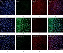
          Epithelial-mesenchymal transition (EMT) is a cellular process in which epithelial cells are partially transformed into stromal cells, which endows the polarized epithelium cells more invasive feature and contributes cancer metastasis and drug resistance. Ferritinophagy is an event of ferritin degradation in lysosomes, which contributes Fenton-mediated ROS production. In addition, some studies have shown that ROS participates in EMT process, but the effect of ROS stemmed from ferritin degradation on EMT has not been fully established. A novel iron chelator, DpdtC (2,2′-di-pyridylketone dithiocarbamate), which could induce ferritinophagy in HepG2 cell in our previous study, was used to investigate its effect on EMT in gastric cancer cells. The proliferation assay showed that DpdtC treatment resulted in growth inhibition and morphologic alteration in MGC-803 cell (IC 50 = 3.1 ± 0.3  μM), and its action involved ROS production that was due to the occurrence of ferritinophagy. More interestingly, DpdtC could also inhibit EMT, leading to the upregulation of E-cadherin and the downregulation of vimentin; however, the addition of NAC and 3-MA could attenuate (or neutralize) the action of DpdtC on ferritinophagy induction and EMT inhibition, supporting that the enhanced ferritinophagic flux contributed to the EMT inhibition. Since the degradation of ferritin may trigger the production of ROS and induce the response of p53, we next studied the role of p53 in the above two-cell events. As expected, an upregulation of p53 was observed after DpdtC insulting; however, the addition of a p53 inhibitor, PFT- α, could significantly attenuate the action of DpdtC on ferritinophagy induction and EMT inhibition. In addition, autophagy inhibitors or NAC could counteract the effect of DpdtC and restore the level of p53 to the control group, indicating that the upregulation of p53 was caused by ferritinophagy-mediated ROS production. In conclusion, our data demonstrated that the inhibition of EMT induced by DpdtC was realized through ferritinophagy-mediated ROS/p53 pathway, which supported that the activation of ferritinophagic flux was the main driving force in EMT inhibition in gastric cancer cells, and further strengthening the concept that NCOA4 participates in EMT process.

          Related collections

          Most cited references42

          • Record: found
          • Abstract: found
          • Article: found

          Comprehensive and Integrative Genomic Characterization of Hepatocellular Carcinoma

          (2017)
          Liver cancer has the second highest worldwide cancer mortality rate and has limited therapeutic options. We analyzed 363 hepatocellular carcinoma (HCC) cases by whole exome sequencing and DNA copy number analyses, and 196 HCC also by DNA methylation, RNA, miRNA, and proteomic expression. DNA sequencing and mutation analysis identified significantly mutated genes including LZTR1 , EEF1A1 , SF3B1 , and SMARCA4 . Significant alterations by mutation or down-regulation by hypermethylation in genes likely to result in HCC metabolic reprogramming ( ALB , APOB , and CPS1 ) were observed. Integrative molecular HCC subtyping incorporating unsupervised clustering of five data platforms identified three subtypes, one of which was associated with poorer prognosis in three HCC cohorts. Integrated analyses enabled development of a p53 target gene expression signature correlating with poor survival. Potential therapeutic targets for which inhibitors exist include WNT signaling, MDM4, MET, VEGFA, MCL1, IDH1, TERT, and immune checkpoint proteins CTLA-4, PD-1, and PD-L1. Multiplex molecular profiling of human hepatocellular carcinoma patients provides insight into subtype characteristics and points toward key pathways to target therapeutically.
            Bookmark
            • Record: found
            • Abstract: found
            • Article: not found

            Mdm2 promotes the rapid degradation of p53.

            The p53 tumour-suppressor protein exerts antiproliferative effects, including growth arrest and apoptosis, in response to various types of stress. The activity of p53 is abrogated by mutations that occur frequently in tumours, as well as by several viral and cellular proteins. The Mdm2 oncoprotein is a potent inhibitor of p53. Mdm2 binds the transcriptional activation domain of p53 and blocks its ability to regulate target genes and to exert antiproliferative effects. On the other hand, p53 activates the expression of the mdm2 gene in an autoregulatory feedback loop. The interval between p53 activation and consequent Mdm2 accumulation defines a time window during which p53 exerts its effects. We now report that Mdm2 also promotes the rapid degradation of p53 under conditions in which p53 is otherwise stabilized. This effect of Mdm2 requires binding of p53; moreover, a small domain of p53, encompassing the Mdm2-binding site, confers Mdm2-dependent detstabilization upon heterologous proteins. Raised amounts of Mdm2 strongly repress mutant p53 accumulation in tumour-derived cells. During recovery from DNA damage, maximal Mdm2 induction coincides with rapid p53 loss. We propose that the Mdm2-promoted degradation of p53 provides a new mechanism to ensure effective termination of the p53 signal.
              Bookmark
              • Record: found
              • Abstract: found
              • Article: not found

              Quantitative proteomics identifies NCOA4 as the cargo receptor mediating ferritinophagy

              Autophagy, the process by which proteins and organelles are sequestered in double-membrane structures called autophagosomes and delivered to lysosomes for degradation, is critical in diseases such as cancer and neurodegeneration 1,2 . Much of our understanding of this process has emerged from analysis of bulk cytoplasmic autophagy, but our understanding of how specific cargo including organelles, proteins, or intracellular pathogens are targeted for selective autophagy is limited 3 . We employed quantitative proteomics to identify a cohort of novel and known autophagosome-enriched proteins, including cargo receptors. Like known cargo receptors, NCOA4 was highly enriched in autophagosomes, and associated with ATG8 proteins that recruit cargo-receptor complexes into autophagosomes. Unbiased identification of NCOA4-associated proteins revealed ferritin heavy and light chains, components of an iron-filled cage structure that protects cells from reactive iron species 4 but is degraded via autophagy to release iron 5,6 through an unknown mechanism. We found that delivery of ferritin to lysosomes required NCOA4, and an inability of NCOA4-deficient cells to degrade ferritin leads to decreased bioavailable intracellular iron. This work identifies NCOA4 as a selective cargo receptor for autophagic turnover of ferritin (ferritinophagy) critical for iron homeostasis and provides a resource for further dissection of autophagosomal cargo-receptor connectivity.
                Bookmark

                Author and article information

                Contributors
                Journal
                Oxid Med Cell Longev
                Oxid Med Cell Longev
                OMCL
                Oxidative Medicine and Cellular Longevity
                Hindawi
                1942-0900
                1942-0994
                2020
                12 March 2020
                : 2020
                : 9762390
                Affiliations
                1Departement of Molecular Biology and Biochemistry, Xinxiang Medical University, Xinxiang, Henan, China 453003
                2Experimental Teaching Center of Biology and Basic Medical Sciences, Sanquan College of Xinxiang Medical University, Xinxiang, Henan, China 453003
                3Department of Histology and Embryology, Sanquan College of Xinxiang Medical University, Xinxiang, Henan, China 453003
                4Laboratory of Molecular Medicine, Xinxiang Medical University, Xinxiang, Henan, China 453003
                Author notes

                Guest Editor: Teruo Miyazaki

                Author information
                https://orcid.org/0000-0001-7293-5313
                https://orcid.org/0000-0002-6226-5843
                Article
                10.1155/2020/9762390
                7091554
                32256964
                ce117618-05f6-480e-8826-a7ba28dd36c8
                Copyright © 2020 Jiankang Feng et al.

                This is an open access article distributed under the Creative Commons Attribution License, which permits unrestricted use, distribution, and reproduction in any medium, provided the original work is properly cited.

                History
                : 24 November 2019
                : 14 February 2020
                Funding
                Funded by: Xinxiang Medical University
                Award ID: SQTD201902
                Award ID: SQTD201703
                Funded by: The Key Research Project Funding Program of Higher Educational Institutions of Henan Province
                Award ID: 19A310021
                Funded by: Henan Science and Technology Agency
                Award ID: 152300410118
                Funded by: National Natural Science Foundation of China
                Award ID: 21571153
                Categories
                Research Article

                Molecular medicine
                Molecular medicine

                Comments

                Comment on this article