Inviting an author to review:
Find an author and click ‘Invite to review selected article’ near their name.
Search for authorsSearch for similar articles
3
views
0
recommends
+1 Recommend
0 collections
    0
    shares
      • Record: found
      • Abstract: found
      • Article: found
      Is Open Access

      Lysosomal integral membrane protein-2 (LIMP-2/SCARB2) is involved in lysosomal cholesterol export

      research-article

      Read this article at

      Bookmark
          There is no author summary for this article yet. Authors can add summaries to their articles on ScienceOpen to make them more accessible to a non-specialist audience.

          Abstract

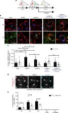
          The intracellular transport of cholesterol is subject to tight regulation. The structure of the lysosomal integral membrane protein type 2 (LIMP-2, also known as SCARB2) reveals a large cavity that traverses the molecule and resembles the cavity in SR-B1 that mediates lipid transfer. The detection of cholesterol within the LIMP-2 structure and the formation of cholesterol like inclusions in LIMP-2 knockout mice suggested the possibility that LIMP2 transports cholesterol in lysosomes. We present results of molecular modeling, crosslinking studies, microscale thermophoresis and cell-based assays that support a role of LIMP-2 in cholesterol transport. We show that the cavity in the luminal domain of LIMP-2 can bind and deliver exogenous cholesterol to the lysosomal membrane and later to lipid droplets. Depletion of LIMP-2 alters SREBP-2-mediated cholesterol regulation, as well as LDL-receptor levels. Our data indicate that LIMP-2 operates in parallel with Niemann Pick (NPC)-proteins, mediating a slower mode of lysosomal cholesterol export.

          Abstract

          Cholesterol transport is tightly regulated in the cell and in lysosomes is regulated by NPC1/2. Here, Heybrock et al. use molecular modeling, knockout mice and cell based studies to show that LIMP-2 also mediates lysosomal cholesterol transport.

          Related collections

          Most cited references30

          • Record: found
          • Abstract: found
          • Article: not found

          Scavenger receptor B2 is a cellular receptor for enterovirus 71.

          Enterovirus 71 (EV71) belongs to human enterovirus species A of the genus Enterovirus within the family Picornaviridae. EV71, together with coxsackievirus A16 (CVA16), are most frequently associated with hand, foot and mouth disease (HFMD). Although HFMD is considered a mild exanthematous infection, infections involving EV71, but not CVA16, can progress to severe neurological disease, including fatal encephalitis, aseptic meningitis and acute flaccid paralysis. In recent years, epidemic and sporadic outbreaks of neurovirulent EV71 infections have been reported in Taiwan, Malaysia, Singapore, Japan and China. Here, we show that human scavenger receptor class B, member 2 (SCARB2, also known as lysosomal integral membrane protein II or CD36b like-2) is a receptor for EV71. EV71 binds soluble SCARB2 or cells expressing SCARB2, and the binding is inhibited by an antibody to SCARB2. Expression of human SCARB2 enables normally unsusceptible cell lines to support EV71 propagation and develop cytopathic effects. EV71 infection is hampered by the antibody to SCARB2 and soluble SCARB2. SCARB2 also supports the infection of the milder pathogen CVA16. The identification of SCARB2 as an EV71 and CVA16 receptor contributes to a better understanding of the pathogenicity of these viruses.
            Bookmark
            • Record: found
            • Abstract: found
            • Article: not found

            Expanding roles for lipid droplets.

            Lipid droplets are the intracellular sites for neutral lipid storage. They are critical for lipid metabolism and energy homeostasis, and their dysfunction has been linked to many diseases. Accumulating evidence suggests that the roles lipid droplets play in biology are significantly broader than previously anticipated. Lipid droplets are the source of molecules important in the nucleus: they can sequester transcription factors and chromatin components and generate the lipid ligands for certain nuclear receptors. Lipid droplets have also emerged as important nodes for fatty acid trafficking, both inside the cell and between cells. In immunity, new roles for droplets, not directly linked to lipid metabolism, have been uncovered, with evidence that they act as assembly platforms for specific viruses and as reservoirs for proteins that fight intracellular pathogens. Until recently, knowledge about droplets in the nervous system has been minimal, but now there are multiple links between lipid droplets and neurodegeneration: many candidate genes for hereditary spastic paraplegia also have central roles in lipid-droplet formation and maintenance, and mitochondrial dysfunction in neurons can lead to transient accumulation of lipid droplets in neighboring glial cells, an event that may, in turn, contribute to neuronal damage. As the cell biology and biochemistry of lipid droplets become increasingly well understood, the next few years should yield many new mechanistic insights into these novel functions of lipid droplets.
              Bookmark
              • Record: found
              • Abstract: found
              • Article: not found

              Retrospective on Cholesterol Homeostasis: The Central Role of Scap.

              Scap is a polytopic membrane protein that functions as a molecular machine to control the cholesterol content of membranes in mammalian cells. In the 21 years since our laboratory discovered Scap, we have learned how it binds sterol regulatory element-binding proteins (SREBPs) and transports them from the endoplasmic reticulum (ER) to the Golgi for proteolytic processing. Proteolysis releases the SREBP transcription factor domains, which enter the nucleus to promote cholesterol synthesis and uptake. When cholesterol in ER membranes exceeds a threshold, the sterol binds to Scap, triggering several conformational changes that prevent the Scap-SREBP complex from leaving the ER. As a result, SREBPs are no longer processed, cholesterol synthesis and uptake are repressed, and cholesterol homeostasis is restored. This review focuses on the four domains of Scap that undergo concerted conformational changes in response to cholesterol binding. The data provide a molecular mechanism for the control of lipids in cell membranes. Expected final online publication date for the Annual Review of Biochemistry Volume 87 is June 20, 2018. Please see http://www.annualreviews.org/page/journal/pubdates for revised estimates.
                Bookmark

                Author and article information

                Contributors
                sergio.grinstein@sickkids.ca
                elina.ikonen@helsinki.fi
                psaftig@biochem.uni-kiel.de
                dneculai@zju.edu.cn
                Journal
                Nat Commun
                Nat Commun
                Nature Communications
                Nature Publishing Group UK (London )
                2041-1723
                6 August 2019
                6 August 2019
                2019
                : 10
                : 3521
                Affiliations
                [1 ]ISNI 0000 0001 2153 9986, GRID grid.9764.c, Biochemisches Institut, , Christian-Albrechts-Universität Kiel, ; Kiel, Germany
                [2 ]ISNI 0000 0004 0410 2071, GRID grid.7737.4, Faculty of Medicine, Anatomy and Stem Cells and Metabolism Research Program, , University of Helsinki, ; Helsinki, Finland
                [3 ]GRID grid.452540.2, Minerva Foundation Institute for Medical Research, ; Helsinki, Finland
                [4 ]ISNI 0000 0004 1759 700X, GRID grid.13402.34, Department of Cell Biology, and Department of Pathology Sir Run Run Shaw Hospital, , Zhejiang University School of Medicine, ; Hangzhou, Zhejiang P.R. China
                [5 ]ISNI 0000 0004 0473 9646, GRID grid.42327.30, Program in Molecular Medicine, , Research Institute, The Hospital for Sick Children, ; Toronto, Ontario M5G 0A4 Canada
                [6 ]ISNI 0000 0001 2157 2938, GRID grid.17063.33, Department of Biochemistry, , University of Toronto, ; Toronto, M5S 1A8 Canada
                [7 ]ISNI 0000 0001 2188 3760, GRID grid.262273.0, Department of Chemistry and Biochemistry, Queens College, , City University of New York, ; Flushing, New York USA
                [8 ]ISNI 0000 0004 0473 9646, GRID grid.42327.30, Cell Biology Program, , Hospital for Sick Children, ; Toronto, M5G 1X8 Canada
                [9 ]ISNI 0000 0001 2157 2938, GRID grid.17063.33, Department of Physiology, , University of Toronto, ; Toronto, M5S 1A8 Canada
                [10 ]Princes Margaret Cancer Centre, Toronto, ON Canada
                [11 ]Laboratory for Membrane Trafficking, VIB-Center for Brain and Disease Research, Leuven, Belgium
                [12 ]ISNI 0000 0001 0944 9128, GRID grid.7491.b, Faculty of Chemistry, Biochemistry III, , University of Bielefeld, ; 33615 Bielefeld, Germany
                [13 ]ISNI 0000 0001 2180 3484, GRID grid.13648.38, Institut für Biochemie und Molekulare Zellbiologie, Zentrum für Experimentelle Medizin, , Universitätsklinikum Hamburg-Eppendorf, ; Hamburg-Eppendorf, Germany
                [14 ]ISNI 0000 0001 2153 9986, GRID grid.9764.c, Institut für Anatomie, , Christian-Albrechts-Universität Kiel, ; Kiel, Germany
                [15 ]ISNI 0000 0001 2299 3507, GRID grid.16753.36, Present Address: Department of Neurology, , Northwestern University Feinberg School of Medicine, ; Chicago, IL 60611 USA
                Author information
                http://orcid.org/0000-0003-1680-9448
                http://orcid.org/0000-0001-6947-5731
                http://orcid.org/0000-0001-9776-5111
                http://orcid.org/0000-0003-3068-9833
                http://orcid.org/0000-0002-0712-4319
                http://orcid.org/0000-0003-0150-9661
                http://orcid.org/0000-0002-5647-1034
                http://orcid.org/0000-0001-8382-1135
                http://orcid.org/0000-0001-8887-9168
                Article
                11425
                10.1038/s41467-019-11425-0
                6684646
                31387993
                cecca28f-3690-40f7-9735-0d56715460ed
                © The Author(s) 2019

                Open Access This article is licensed under a Creative Commons Attribution 4.0 International License, which permits use, sharing, adaptation, distribution and reproduction in any medium or format, as long as you give appropriate credit to the original author(s) and the source, provide a link to the Creative Commons license, and indicate if changes were made. The images or other third party material in this article are included in the article’s Creative Commons license, unless indicated otherwise in a credit line to the material. If material is not included in the article’s Creative Commons license and your intended use is not permitted by statutory regulation or exceeds the permitted use, you will need to obtain permission directly from the copyright holder. To view a copy of this license, visit http://creativecommons.org/licenses/by/4.0/.

                History
                : 12 September 2018
                : 12 July 2019
                Funding
                Funded by: FundRef https://doi.org/10.13039/501100001659, Deutsche Forschungsgemeinschaft (German Research Foundation);
                Award ID: GRK1459
                Award ID: FOR2625
                Award Recipient :
                Funded by: FundRef https://doi.org/10.13039/501100002341, Academy of Finland (Suomen Akatemia);
                Award ID: 282192
                Award Recipient :
                Funded by: Key Program of Zhejiang Provincial Natural Science Foundation of China, Finnish Cultural Foundation, Häme Regional Fund and University of Helsinki, Canadian Institutes of Health Research, VIB, FWO
                Categories
                Article
                Custom metadata
                © The Author(s) 2019

                Uncategorized
                sterols,lysosomes
                Uncategorized
                sterols, lysosomes

                Comments

                Comment on this article