17
views
0
recommends
+1 Recommend
1 collections
    0
    shares
      • Record: found
      • Abstract: found
      • Article: not found

      Hydroxychloroquine as Postexposure Prophylaxis to Prevent Severe Acute Respiratory Syndrome Coronavirus 2 Infection : A Randomized Trial

      other
      , MBChB, MSc, DPhil, , ScD, , PhD, , MD, , MD, MPH, , PhD, MPH, , MD, , MD, MPH, , MD, MPH,   , MD, PhD, , MD, , PhD, MPH, , MD, , MD, , MD, MSc, , MD, , MS, , BA, , MS, , MD, MPH, , MS, , MPH, , MD, , MD, PhD, MS, MPhil, , PhD, , MD, PhD, , MD, MPH, , MD, MPH, , MD, MPH, , MD, PhD * , for the Hydroxychloroquine COVID-19 PEP Study Team
      Annals of Internal Medicine
      American College of Physicians

      Read this article at

      ScienceOpenPublisherPMC
      Bookmark
          There is no author summary for this article yet. Authors can add summaries to their articles on ScienceOpen to make them more accessible to a non-specialist audience.

          Abstract

          Clinical trials have ruled out a role of hydroxychloroquine in the treatment of COVID-19, but it has been hypothesized that hydroxychloroquine's activity against SARS-CoV-2 in the laboratory suggests a role in prevention. This randomized controlled trial tests hydroxychloroquine as postexposure prophylaxis for SARS-CoV-2 infection.

          Abstract

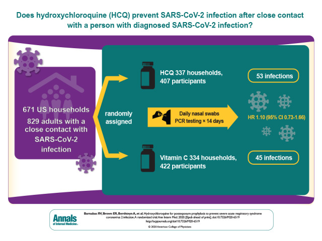
          Visual Abstract. Postexposure Hydroxychloroquine Prophylaxis to Prevent SARS-CoV-2 Infection  Clinical trials have ruled out a role of hydroxychloroquine in the treatment of COVID-19, but it has been hypothesized that hydroxychloroquine's activity against SARS-CoV-2 in the laboratory suggests a role in prevention. This randomized controlled trial tests hydroxychloroquine as postexposure prophylaxis for SARS-CoV-2 infection.
          Visual Abstract.
          Postexposure Hydroxychloroquine Prophylaxis to Prevent SARS-CoV-2 Infection

          Clinical trials have ruled out a role of hydroxychloroquine in the treatment of COVID-19, but it has been hypothesized that hydroxychloroquine's activity against SARS-CoV-2 in the laboratory suggests a role in prevention. This randomized controlled trial tests hydroxychloroquine as postexposure prophylaxis for SARS-CoV-2 infection.

          Abstract

          Background:

          Effective prevention against coronavirus disease 2019 (COVID-19), caused by severe acute respiratory syndrome coronavirus 2 (SARS-CoV-2), is currently limited to nonpharmaceutical strategies. Laboratory and observational data suggested that hydroxychloroquine had biological activity against SARS-CoV-2, potentially permitting its use for prevention.

          Objective:

          To test hydroxychloroquine as postexposure prophylaxis for SARS-CoV-2 infection.

          Design:

          Household-randomized, double-blind, controlled trial of hydroxychloroquine postexposure prophylaxis. (ClinicalTrials.gov: NCT04328961)

          Setting:

          National U.S. multicenter study.

          Participants:

          Close contacts recently exposed (<96 hours) to persons with diagnosed SARS-CoV-2 infection.

          Intervention:

          Hydroxychloroquine (400 mg/d for 3 days followed by 200 mg/d for 11 days) or ascorbic acid (500 mg/d followed by 250 mg/d) as a placebo-equivalent control.

          Measurements:

          Participants self-collected mid-turbinate swabs daily (days 1 to 14) for SARS-CoV-2 polymerase chain reaction (PCR) testing. The primary outcome was PCR-confirmed incident SARS-CoV-2 infection among persons who were SARS-CoV-2 negative at enrollment.

          Results:

          Between March and August 2020, 671 households were randomly assigned: 337 (407 participants) to the hydroxychloroquine group and 334 (422 participants) to the control group. Retention at day 14 was 91%, and 10 724 of 11 606 (92%) expected swabs were tested. Among the 689 (89%) participants who were SARS-CoV-2 negative at baseline, there was no difference between the hydroxychloroquine and control groups in SARS-CoV-2 acquisition by day 14 (53 versus 45 events; adjusted hazard ratio, 1.10 [95% CI, 0.73 to 1.66]; P > 0.20). The frequency of participants experiencing adverse events was higher in the hydroxychloroquine group than the control group (66 [16.2%] versus 46 [10.9%], respectively; P = 0.026).

          Limitation:

          The delay between exposure, and then baseline testing and the first dose of hydroxychloroquine or ascorbic acid, was a median of 2 days.

          Conclusion:

          This rigorous randomized controlled trial among persons with recent exposure excluded a clinically meaningful effect of hydroxychloroquine as postexposure prophylaxis to prevent SARS-CoV-2 infection.

          Primary Funding Source:

          Bill & Melinda Gates Foundation.

          Related collections

          Most cited references26

          • Record: found
          • Abstract: found
          • Article: found

          Characteristics of and Important Lessons From the Coronavirus Disease 2019 (COVID-19) Outbreak in China: Summary of a Report of 72 314 Cases From the Chinese Center for Disease Control and Prevention

            Bookmark
            • Record: found
            • Abstract: found
            • Article: not found

            An interactive web-based dashboard to track COVID-19 in real time

            In December, 2019, a local outbreak of pneumonia of initially unknown cause was detected in Wuhan (Hubei, China), and was quickly determined to be caused by a novel coronavirus, 1 namely severe acute respiratory syndrome coronavirus 2 (SARS-CoV-2). The outbreak has since spread to every province of mainland China as well as 27 other countries and regions, with more than 70 000 confirmed cases as of Feb 17, 2020. 2 In response to this ongoing public health emergency, we developed an online interactive dashboard, hosted by the Center for Systems Science and Engineering (CSSE) at Johns Hopkins University, Baltimore, MD, USA, to visualise and track reported cases of coronavirus disease 2019 (COVID-19) in real time. The dashboard, first shared publicly on Jan 22, illustrates the location and number of confirmed COVID-19 cases, deaths, and recoveries for all affected countries. It was developed to provide researchers, public health authorities, and the general public with a user-friendly tool to track the outbreak as it unfolds. All data collected and displayed are made freely available, initially through Google Sheets and now through a GitHub repository, along with the feature layers of the dashboard, which are now included in the Esri Living Atlas. The dashboard reports cases at the province level in China; at the city level in the USA, Australia, and Canada; and at the country level otherwise. During Jan 22–31, all data collection and processing were done manually, and updates were typically done twice a day, morning and night (US Eastern Time). As the outbreak evolved, the manual reporting process became unsustainable; therefore, on Feb 1, we adopted a semi-automated living data stream strategy. Our primary data source is DXY, an online platform run by members of the Chinese medical community, which aggregates local media and government reports to provide cumulative totals of COVID-19 cases in near real time at the province level in China and at the country level otherwise. Every 15 min, the cumulative case counts are updated from DXY for all provinces in China and for other affected countries and regions. For countries and regions outside mainland China (including Hong Kong, Macau, and Taiwan), we found DXY cumulative case counts to frequently lag behind other sources; we therefore manually update these case numbers throughout the day when new cases are identified. To identify new cases, we monitor various Twitter feeds, online news services, and direct communication sent through the dashboard. Before manually updating the dashboard, we confirm the case numbers with regional and local health departments, including the respective centres for disease control and prevention (CDC) of China, Taiwan, and Europe, the Hong Kong Department of Health, the Macau Government, and WHO, as well as city-level and state-level health authorities. For city-level case reports in the USA, Australia, and Canada, which we began reporting on Feb 1, we rely on the US CDC, the government of Canada, the Australian Government Department of Health, and various state or territory health authorities. All manual updates (for countries and regions outside mainland China) are coordinated by a team at Johns Hopkins University. The case data reported on the dashboard aligns with the daily Chinese CDC 3 and WHO situation reports 2 for within and outside of mainland China, respectively (figure ). Furthermore, the dashboard is particularly effective at capturing the timing of the first reported case of COVID-19 in new countries or regions (appendix). With the exception of Australia, Hong Kong, and Italy, the CSSE at Johns Hopkins University has reported newly infected countries ahead of WHO, with Hong Kong and Italy reported within hours of the corresponding WHO situation report. Figure Comparison of COVID-19 case reporting from different sources Daily cumulative case numbers (starting Jan 22, 2020) reported by the Johns Hopkins University Center for Systems Science and Engineering (CSSE), WHO situation reports, and the Chinese Center for Disease Control and Prevention (Chinese CDC) for within (A) and outside (B) mainland China. Given the popularity and impact of the dashboard to date, we plan to continue hosting and managing the tool throughout the entirety of the COVID-19 outbreak and to build out its capabilities to establish a standing tool to monitor and report on future outbreaks. We believe our efforts are crucial to help inform modelling efforts and control measures during the earliest stages of the outbreak.
              Bookmark
              • Record: found
              • Abstract: found
              • Article: found
              Is Open Access

              Remdesivir and chloroquine effectively inhibit the recently emerged novel coronavirus (2019-nCoV) in vitro

              Dear Editor, In December 2019, a novel pneumonia caused by a previously unknown pathogen emerged in Wuhan, a city of 11 million people in central China. The initial cases were linked to exposures in a seafood market in Wuhan. 1 As of January 27, 2020, the Chinese authorities reported 2835 confirmed cases in mainland China, including 81 deaths. Additionally, 19 confirmed cases were identified in Hong Kong, Macao and Taiwan, and 39 imported cases were identified in Thailand, Japan, South Korea, United States, Vietnam, Singapore, Nepal, France, Australia and Canada. The pathogen was soon identified as a novel coronavirus (2019-nCoV), which is closely related to sever acute respiratory syndrome CoV (SARS-CoV). 2 Currently, there is no specific treatment against the new virus. Therefore, identifying effective antiviral agents to combat the disease is urgently needed. An efficient approach to drug discovery is to test whether the existing antiviral drugs are effective in treating related viral infections. The 2019-nCoV belongs to Betacoronavirus which also contains SARS-CoV and Middle East respiratory syndrome CoV (MERS-CoV). Several drugs, such as ribavirin, interferon, lopinavir-ritonavir, corticosteroids, have been used in patients with SARS or MERS, although the efficacy of some drugs remains controversial. 3 In this study, we evaluated the antiviral efficiency of five FAD-approved drugs including ribavirin, penciclovir, nitazoxanide, nafamostat, chloroquine and two well-known broad-spectrum antiviral drugs remdesivir (GS-5734) and favipiravir (T-705) against a clinical isolate of 2019-nCoV in vitro. Standard assays were carried out to measure the effects of these compounds on the cytotoxicity, virus yield and infection rates of 2019-nCoVs. Firstly, the cytotoxicity of the candidate compounds in Vero E6 cells (ATCC-1586) was determined by the CCK8 assay. Then, Vero E6 cells were infected with nCoV-2019BetaCoV/Wuhan/WIV04/2019 2 at a multiplicity of infection (MOI) of 0.05 in the presence of varying concentrations of the test drugs. DMSO was used in the controls. Efficacies were evaluated by quantification of viral copy numbers in the cell supernatant via quantitative real-time RT-PCR (qRT-PCR) and confirmed with visualization of virus nucleoprotein (NP) expression through immunofluorescence microscopy at 48 h post infection (p.i.) (cytopathic effect was not obvious at this time point of infection). Among the seven tested drugs, high concentrations of three nucleoside analogs including ribavirin (half-maximal effective concentration (EC50) = 109.50 μM, half-cytotoxic concentration (CC50) > 400 μM, selectivity index (SI) > 3.65), penciclovir (EC50 = 95.96 μM, CC50 > 400 μM, SI > 4.17) and favipiravir (EC50 = 61.88 μM, CC50 > 400 μM, SI > 6.46) were required to reduce the viral infection (Fig. 1a and Supplementary information, Fig. S1). However, favipiravir has been shown to be 100% effective in protecting mice against Ebola virus challenge, although its EC50 value in Vero E6 cells was as high as 67 μM, 4 suggesting further in vivo studies are recommended to evaluate this antiviral nucleoside. Nafamostat, a potent inhibitor of MERS-CoV, which prevents membrane fusion, was inhibitive against the 2019-nCoV infection (EC50 = 22.50 μM, CC50 > 100 μM, SI > 4.44). Nitazoxanide, a commercial antiprotozoal agent with an antiviral potential against a broad range of viruses including human and animal coronaviruses, inhibited the 2019-nCoV at a low-micromolar concentration (EC50 = 2.12 μM; CC50 > 35.53 μM; SI > 16.76). Further in vivo evaluation of this drug against 2019-nCoV infection is recommended. Notably, two compounds remdesivir (EC50 = 0.77 μM; CC50 > 100 μM; SI > 129.87) and chloroquine (EC50 = 1.13 μM; CC50 > 100 μM, SI > 88.50) potently blocked virus infection at low-micromolar concentration and showed high SI (Fig. 1a, b). Fig. 1 The antiviral activities of the test drugs against 2019-nCoV in vitro. a Vero E6 cells were infected with 2019-nCoV at an MOI of 0.05 in the treatment of different doses of the indicated antivirals for 48 h. The viral yield in the cell supernatant was then quantified by qRT-PCR. Cytotoxicity of these drugs to Vero E6 cells was measured by CCK-8 assays. The left and right Y-axis of the graphs represent mean % inhibition of virus yield and cytotoxicity of the drugs, respectively. The experiments were done in triplicates. b Immunofluorescence microscopy of virus infection upon treatment of remdesivir and chloroquine. Virus infection and drug treatment were performed as mentioned above. At 48 h p.i., the infected cells were fixed, and then probed with rabbit sera against the NP of a bat SARS-related CoV 2 as the primary antibody and Alexa 488-labeled goat anti-rabbit IgG (1:500; Abcam) as the secondary antibody, respectively. The nuclei were stained with Hoechst dye. Bars, 100 μm. c and d Time-of-addition experiment of remdesivir and chloroquine. For “Full-time” treatment, Vero E6 cells were pre-treated with the drugs for 1 h, and virus was then added to allow attachment for 2 h. Afterwards, the virus–drug mixture was removed, and the cells were cultured with drug-containing medium until the end of the experiment. For “Entry” treatment, the drugs were added to the cells for 1 h before viral attachment, and at 2 h p.i., the virus–drug mixture was replaced with fresh culture medium and maintained till the end of the experiment. For “Post-entry” experiment, drugs were added at 2 h p.i., and maintained until the end of the experiment. For all the experimental groups, cells were infected with 2019-nCoV at an MOI of 0.05, and virus yield in the infected cell supernatants was quantified by qRT-PCR c and NP expression in infected cells was analyzed by Western blot d at 14 h p.i. Remdesivir has been recently recognized as a promising antiviral drug against a wide array of RNA viruses (including SARS/MERS-CoV 5 ) infection in cultured cells, mice and nonhuman primate (NHP) models. It is currently under clinical development for the treatment of Ebola virus infection. 6 Remdesivir is an adenosine analogue, which incorporates into nascent viral RNA chains and results in pre-mature termination. 7 Our time-of-addition assay showed remdesivir functioned at a stage post virus entry (Fig. 1c, d), which is in agreement with its putative anti-viral mechanism as a nucleotide analogue. Warren et al. showed that in NHP model, intravenous administration of 10 mg/kg dose of remdesivir resulted in concomitant persistent levels of its active form in the blood (10 μM) and conferred 100% protection against Ebola virus infection. 7 Our data showed that EC90 value of remdesivir against 2019-nCoV in Vero E6 cells was 1.76 μM, suggesting its working concentration is likely to be achieved in NHP. Our preliminary data (Supplementary information, Fig. S2) showed that remdesivir also inhibited virus infection efficiently in a human cell line (human liver cancer Huh-7 cells), which is sensitive to 2019-nCoV. 2 Chloroquine, a widely-used anti-malarial and autoimmune disease drug, has recently been reported as a potential broad-spectrum antiviral drug. 8,9 Chloroquine is known to block virus infection by increasing endosomal pH required for virus/cell fusion, as well as interfering with the glycosylation of cellular receptors of SARS-CoV. 10 Our time-of-addition assay demonstrated that chloroquine functioned at both entry, and at post-entry stages of the 2019-nCoV infection in Vero E6 cells (Fig. 1c, d). Besides its antiviral activity, chloroquine has an immune-modulating activity, which may synergistically enhance its antiviral effect in vivo. Chloroquine is widely distributed in the whole body, including lung, after oral administration. The EC90 value of chloroquine against the 2019-nCoV in Vero E6 cells was 6.90 μM, which can be clinically achievable as demonstrated in the plasma of rheumatoid arthritis patients who received 500 mg administration. 11 Chloroquine is a cheap and a safe drug that has been used for more than 70 years and, therefore, it is potentially clinically applicable against the 2019-nCoV. Our findings reveal that remdesivir and chloroquine are highly effective in the control of 2019-nCoV infection in vitro. Since these compounds have been used in human patients with a safety track record and shown to be effective against various ailments, we suggest that they should be assessed in human patients suffering from the novel coronavirus disease. Supplementary information Supplementary information, Materials and Figures
                Bookmark

                Author and article information

                Journal
                Ann Intern Med
                Ann Intern Med
                aim
                Annals of Internal Medicine
                American College of Physicians
                0003-4819
                1539-3704
                8 December 2020
                : M20-6519
                Affiliations
                [1 ]University of Washington and Fred Hutchinson Cancer Research Center, Seattle, Washington (R.V.B., E.R.B., C.J., A.L.G., K.R.J., A.W.)
                [2 ]New York University Grossman School of Medicine, New York, New York (A.B., L.E.T., A.K.)
                [3 ]University of Washington, Seattle, Washington (H.C.S., T.T.S., M.L.K., K.K.T., S.M., H.S.H., L.K., M.W., C.C., H.Y.C., J.M.B.)
                [4 ]University of Maryland School of Medicine, Baltimore, Maryland (K.M.N., M.K.L., M.D.)
                [5 ]Boston University School of Medicine and Boston Medical Center, Boston, Massachusetts (M.K.P.)
                [6 ]School of Public Health and Tropical Medicine, Tulane University, New Orleans, Louisiana (P.J.K.)
                [7 ]School of Medicine, Tulane University, New Orleans, Louisiana (A.L.)
                [8 ]State University of New York Upstate Medical University, Syracuse, New York (K.P.)
                [9 ]University of California, Los Angeles, California (R.J.L., R.H.)
                [10 ]Fred Hutchinson Cancer Research Center, Seattle, Washington (M.H.)
                Author notes
                Acknowledgment: The authors thank the study participants for their motivation and dedication; the members of the trial's Data and Safety Monitoring Board (Drs. David Glidden, Michael Boeckh, and Robert Coombs), local advisors at each trial site, and overseeing ethics review committees for their expertise and guidance; Drs. Scott Miller and Peter Dull from the Bill & Melinda Gates Foundation for their attentive oversight; and the HCQ COVID-19 PEP Study Team for their dedication and perseverance.
                Disclaimer: The content is solely the responsibility of the authors and does not necessarily represent the views, decisions, or policies of the institutions with which they are affiliated or the HCQ COVID-19 PEP Study funders.
                Funding: The HCQ COVID-19 PEP Study was funded by the Bill & Melinda Gates Foundation (INV-016204) through the COVID-19 Therapeutics Accelerator and the University of Washington King K. Holmes Endowed Professorship in STDs and AIDS. Hydroxychloroquine for the study was donated by Sandoz.
                Reproducible Research Statement: Study protocol: Available at ClinicalTrials.gov (NCT02929992). Statistical code: Statistical code: The Cox model fit call is provided in Supplement Table 1; other file management aspects of the statistical code are not available. Data set: A complete deidentified patient data set sufficient to reproduce the study findings will be made available no later than 1 year after completion of the trial, after approval of a concept sheet summarizing the analyses to be done. Further inquiries can be directed to the HCQ COVID-19 PEP Study Scientific Committee (e-mail, icrc@ 123456uw.edu ).
                Corresponding Author: Ruanne V. Barnabas, International Clinical Research Center (ICRC), Department of Global Health, University of Washington, UW Box 359927, 325 Ninth Avenue, Seattle, WA 98104; e-mail, rbarnaba@ 123456uw.edu .
                Current Author Addresses: Drs. Barnabas, Stankiewicz Karita, Morrison, Celum, and Baeten; Mr. Schaafsma; Ms. Krows; Ms. Thomas; Mr. Haugen; and Ms. Kidoguchi: International Clinical Research Center (ICRC), Department of Global Health, University of Washington, UW Box 359927, 325 Ninth Avenue, Seattle, WA 98104.
                Dr. Brown: Fred Hutchinson Cancer Research Center, 1100 Fairview Avenue North, Seattle, WA 98109.
                Dr. Bershteyn: Translational Research Building, 227 East 30th Street, New York, NY 10016.
                Dr. Johnston: International Clinical Research Center (ICRC), Department of Global Health, University of Washington, UW Box 359928, 325 Ninth Avenue, Seattle, WA 98104.
                Dr. Thorpe: NYU Grossman School of Medicine, 180 Madison Avenue, New York, NY 10016.
                Dr. Kottkamp: Bellevue Hospital, 462 First Avenue, H Building, 16S 5-13, New York, NY 10016.
                Drs. Neuzil, Laufer, and Deming: Center for Vaccine Development and Global Health, University of Maryland School of Medicine, 685 West Baltimore Street, Room 480, Baltimore, MD 21201.
                Dr. Paasche-Orlow: Boston Medical Center, 801 Massachusetts Avenue, Second Floor, Boston, MA 02119.
                Dr. Kissinger: Tulane University School of Public Health and Tropical Medicine, 1440 Canal Street, Suite 2004, New Orleans, LA 70112.
                Dr. Luk: Tulane University Health Sciences Center, Section of Infectious Disease, 1415 Tulane Avenue, New Orleans, LA 70112.
                Dr. Paolino: Upstate Medical University, Infectious Disease Division, 725 Irving Avenue, Suite 311, Syracuse, NY 13210.
                Dr. Landovitz: UCLA Center for Clinical AIDS Research & Education, 911 Broxton Avenue, Suite 200, Los Angeles, CA 90024.
                Dr. Hoffman: UCLA Department of Medicine, Division of Infectious Diseases, 10833 Le Conte Avenue, 52-215 CHS, Los Angeles, CA 90095.
                Dr. Wener: University of Washington Medical Center, Department of Laboratory Medicine and Pathology, Box 357110, Seattle, WA 98195.
                Drs. Greninger and Huang: University of Washington, Virology Laboratory, 1616 Eastlake Avenue East, Suite 320, Seattle, WA 98102.
                Dr. Jerome: Fred Hutchinson Cancer Research Center, 1100 Fairview Avenue North, E5-110, Seattle, WA 98109.
                Dr. Wald: University of Washington, 325 Ninth Avenue, HMC#359928, Seattle, WA 98104.
                Dr. Chu: University of Washington, 750 Republican Street, UWMC#358061, Seattle, WA 98109.
                Author Contributions: Conception and design: R.V. Barnabas, E.R. Brown, A. Bershteyn, K.M. Neuzil, M.L. Krows, M. Wener, A. Wald, C. Celum, H.Y. Chu, J.M. Baeten.
                Analysis and interpretation of the data: R.V. Barnabas, E.R. Brown, C. Johnston, L.E. Thorpe, K.M. Neuzil, M.K. Laufer, P.J. Kissinger, K. Paolino, R.J. Landovitz, R.M. Hoffman, T.T. Schaafsma, K.K. Thomas, L. Kidoguchi, M.L. Huang, K.R. Jerome, A. Wald, C. Celum, H.Y. Chu, J.M. Baeten.
                Drafting of the article: R.V. Barnabas, E.R. Brown, K.M. Neuzil, M.K. Laufer, P.J. Kissinger, T.T. Schaafsma, M.L. Krows, C. Celum.
                Critical revision for important intellectual content: R.V. Barnabas, E.R. Brown, A. Bershteyn, H.C. Stankiewicz Karita, C. Johnston, L.E. Thorpe, A. Kottkamp, K.M. Neuzil, M.K. Laufer, M.K. Paasche-Orlow, P.J. Kissinger, K. Paolino, R.J. Landovitz, R.M. Hoffman, T.T. Schaafsma, K.K. Thomas, S. Morrison, M. Wener, A. Wald, H.Y. Chu, J.M. Baeten.
                Final approval of the article: R.V. Barnabas, E.R. Brown, A. Bershteyn, H.C. Stankiewicz Karita, C. Johnston, L.E. Thorpe, A. Kottkamp, K.M. Neuzil, M.K. Laufer, M. Deming, M.K. Paasche-Orlow, P.J. Kissinger, A. Luk, R.J. Landovitz, K. Paolino, R.M. Hoffman, T.T. Schaafsma, M.L. Krows, K.K. Thomas, S. Morrison, H.S. Haugen, L. Kidoguchi, M. Wener, A.L. Greninger, M.L. Huang, K.R. Jerome, A. Wald, C. Celum, H.Y. Chu, J.M. Baeten.
                Provision of study materials or patients: H.C. Stankiewicz Karita, A. Kottkamp, M.K. Laufer, M. Deming, M.K. Paasche-Orlow, P.J. Kissinger, A. Luk, R.J. Landovitz, R.M. Hoffman, A. Wald.
                Statistical expertise: E.R. Brown, T.T. Schaafsma, K.K. Thomas.
                Obtaining of funding: R.V. Barnabas, J.M. Baeten.
                Administrative, technical, or logistic support: H.C. Stankiewicz Karita, L.E. Thorpe, A. Kottkamp, M.K. Laufer, M.K. Paasche-Orlow, A.L. Greninger, H.S. Haugen, M. Wener, M.L. Huang, K.R. Jerome, A. Wald, J.M. Baeten.
                Collection and assembly of data: A. Bershteyn, H.C. Stankiewicz Karita, C. Johnston, L.E. Thorpe, A Kottkamp, K.M. Neuzil, M.K. Laufer, M.K. Paasche-Orlow, P.J. Kissinger, K. Paolino, R.J. Landovitz, R.M. Hoffman, T.T. Schaafsma, S. Morrison, A.L. Greninger, M.L. Huang, K.R. Jerome, C. Celum, J.M. Baeten.
                Author information
                https://orcid.org/0000-0003-4286-2111
                https://orcid.org/0000-0002-6604-7093
                https://orcid.org/0000-0002-9458-286X
                https://orcid.org/0000-0002-3073-0843
                https://orcid.org/0000-0002-5535-2674
                https://orcid.org/0000-0003-4103-1666
                https://orcid.org/0000-0001-8300-9593
                https://orcid.org/0000-0003-2574-0542
                https://orcid.org/0000-0001-9575-4574
                https://orcid.org/0000-0002-7024-468X
                https://orcid.org/0000-0002-2034-2150
                https://orcid.org/0000-0002-8304-6957
                https://orcid.org/0000-0002-7443-0527
                https://orcid.org/0000-0003-3486-6438
                Article
                aim-olf-M206519
                10.7326/M20-6519
                7732017
                33284679
                cf487630-d9db-4406-9381-ceace1035249
                Copyright @ 2020

                This article is made available via the PMC Open Access Subset for unrestricted re-use for research, analyses, and text and data mining through PubMed Central. Acknowledgement of the original source shall include a notice similar to the following: "© 2020 American College of Physicians. Some rights reserved. This work permits non-commercial use, distribution, and reproduction in any medium, provided the original author and source are credited." These permissions are granted for the duration of the COVID-19 pandemic or until permissions are revoked in writing. Upon expiration of these permissions, PMC is granted a perpetual license to make this article available via PMC and Europe PMC, consistent with existing copyright protections.

                History
                Categories
                Original Research
                early, Currently Online First
                coronavirus, Coronavirus Disease 2019 (COVID-19)
                poc-eligible, POC Eligible
                3122457, COVID-19
                2357, Health care providers
                11279, SARS coronavirus
                9715, Patients
                6354, Upper respiratory tract infections
                1541398, Pulmonary diseases
                3282, Infectious diseases
                8910, Epidemiology
                7245, Lungs

                Comments

                Comment on this article