131
views
0
recommends
+1 Recommend
0 collections
    0
    shares
      • Record: found
      • Abstract: found
      • Article: found
      Is Open Access

      Increasing tPA Activity in Astrocytes Induced by Multipotent Mesenchymal Stromal Cells Facilitate Neurite Outgrowth after Stroke in the Mouse

      research-article

      Read this article at

      Bookmark
          There is no author summary for this article yet. Authors can add summaries to their articles on ScienceOpen to make them more accessible to a non-specialist audience.

          Abstract

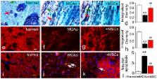
          We demonstrate that tissue plasminogen activator (tPA) and its inhibitors contribute to neurite outgrowth in the central nervous system (CNS) after treatment of stroke with multipotent mesenchymal stromal cells (MSCs). In vivo, administration of MSCs to mice subjected to middle cerebral artery occlusion (MCAo) significantly increased activation of tPA and downregulated PAI-1 levels in the ischemic boundary zone (IBZ) compared with control PBS treated mice, concurrently with increases of myelinated axons and synaptophysin. In vitro, MSCs significantly increased tPA levels and concomitantly reduced plasminogen activator inhibitor 1 (PAI-1) expression in astrocytes under normal and oxygen and glucose deprivation (OGD) conditions. ELISA analysis of conditioned medium revealed that MSCs stimulated astrocytes to secrete tPA. When primary cortical neurons were cultured in the conditioned medium from MSC co-cultured astrocytes, these neurons exhibited a significant increase in neurite outgrowth compared to conditioned medium from astrocytes alone. Blockage of tPA with a neutralizing antibody or knock-down of tPA with siRNA significantly attenuated the effect of the conditioned medium on neurite outgrowth. Addition of recombinant human tPA into cortical neuronal cultures also substantially enhanced neurite outgrowth. Collectively, these in vivo and in vitro data suggest that the MSC mediated increased activation of tPA in astrocytes promotes neurite outgrowth after stroke.

          Related collections

          Most cited references86

          • Record: found
          • Abstract: found
          • Article: not found

          Activation of PDGF-CC by tissue plasminogen activator impairs blood-brain barrier integrity during ischemic stroke.

          Thrombolytic treatment of ischemic stroke with tissue plasminogen activator (tPA) is markedly limited owing to concerns about hemorrhagic complications and the requirement that tPA be administered within 3 h of symptoms. Here we report that tPA activation of latent platelet-derived growth factor-CC (PDGF-CC) may explain these limitations. Intraventricular injection of tPA or active PDGF-CC, in the absence of ischemia, leads to significant increases in cerebrovascular permeability. In contrast, co-injection of neutralizing antibodies to PDGF-CC with tPA blocks this increased permeability, indicating that PDGF-CC is a downstream substrate of tPA within the neurovascular unit. These effects are mediated through activation of PDGF-alpha receptors (PDGFR-alpha) on perivascular astrocytes, and treatment of mice with the PDGFR-alpha antagonist imatinib after ischemic stroke reduces both cerebrovascular permeability and hemorrhagic complications associated with late administration of thrombolytic tPA. These data demonstrate that PDGF signaling regulates blood-brain barrier permeability and suggest potential new strategies for stroke treatment.
            Bookmark
            • Record: found
            • Abstract: found
            • Article: not found

            Protective role of reactive astrocytes in brain ischemia.

            Reactive astrocytes are thought to protect the penumbra during brain ischemia, but direct evidence has been lacking due to the absence of suitable experimental models. Previously, we generated mice deficient in two intermediate filament (IF) proteins, glial fibrillary acidic protein (GFAP) and vimentin, whose upregulation is the hallmark of reactive astrocytes. GFAP(-/-)Vim(-/-) mice exhibit attenuated posttraumatic reactive gliosis, improved integration of neural grafts, and posttraumatic regeneration. Seven days after middle cerebral artery (MCA) transection, infarct volume was 210 to 350% higher in GFAP(-/-)Vim(-/-) than in wild-type (WT) mice; GFAP(-/-), Vim(-/-) and WT mice had the same infarct volume. Endothelin B receptor (ET(B)R) immunoreactivity was strong on cultured astrocytes and reactive astrocytes around infarct in WT mice but undetectable in GFAP(-/-)Vim(-/-) astrocytes. In WT astrocytes, ET(B)R colocalized extensively with bundles of IFs. GFAP(-/-)Vim(-/-) astrocytes showed attenuated endothelin-3-induced blockage of gap junctions. Total and glutamate transporter-1 (GLT-1)-mediated glutamate transport was lower in GFAP(-/-)Vim(-/-) than in WT mice. DNA array analysis and quantitative real-time PCR showed downregulation of plasminogen activator inhibitor-1 (PAI-1), an inhibitor of tissue plasminogen activator. Thus, reactive astrocytes have a protective role in brain ischemia, and the absence of astrocyte IFs is linked to changes in glutamate transport, ET(B)R-mediated control of gap junctions, and PAI-1 expression.
              Bookmark
              • Record: found
              • Abstract: found
              • Article: not found

              Intravenous bone marrow stromal cell therapy reduces apoptosis and promotes endogenous cell proliferation after stroke in female rat.

              The present study investigates the induction of neurogenesis, reduction of apoptosis, and promotion of basic fibroblast growth factor (bFGF) expression as possible mechanisms by which treatment of stroke with bone marrow stromal cells (MSCs) improves neurological functional recovery. Additionally, for the first time, we treated cerebral ischemia in female rats with intraveneous administration of MSCs. Female rats were subjected to 2 hr of middle cerebral artery occlusion (MCAo), followed by an injection of 3 x 10(6) male (for Y chromosome labeling) rat MSCs or phosphate-buffered saline (PBS) into the tail vein 24 hr after MCAo. All animals received daily injection of bromodeoxyuridine (BrdU; 50 mg/kg, i.p.) for 13 days after treatment for identification of newly synthesized DNA. Animals were sacrificed at 14 days after MCAo. Behavioral tests (rotarod and adhesive-removal tests) were performed. In situ hybridization, immunohistochemistry, and terminal deoxynucleotidyltransferase (TdT)-mediated dUTP-biotin nick-end labeling (TUNEL) were performed to identify transplanted MSCs (Y chromosome), BrdU, bFGF, and apoptotic cells in the brain. Significant recovery of behavior was found in MSC-treated rats at 7 days in the somatosensory test and at 14 days in the motor test after MCAo compared with control, PBS-treated animals (P<.05). MSCs were found to survive and preferentially localize to the ipsilateral ischemic hemisphere. Significantly more BrdU-positive cells were located in the subventricular zone (P<.05), and significantly fewer apoptotic cells and more bFGF immunoreactive cell were found in the ischemic boundary area (P<.05) of MSC-treated rats than in PBS-treated animals. Here we demonstrate that intravenously administered male MSCs increase bFGF expression, reduce apoptosis, promote endogenous cellular proliferation, and improve functional recovery after stroke in female rats. Copyright 2003 Wiley-Liss, Inc.
                Bookmark

                Author and article information

                Contributors
                Role: Editor
                Journal
                PLoS One
                plos
                plosone
                PLoS ONE
                Public Library of Science (San Francisco, USA )
                1932-6203
                2010
                3 February 2010
                : 5
                : 2
                : e9027
                Affiliations
                [1 ]Department of Neurology, Henry Ford Health System, Detroit, Michigan, United States of America
                [2 ]Department of Physics, Oakland University, Rochester, Michigan, United States of America
                University of São Paulo, Brazil
                Author notes

                Conceived and designed the experiments: HX YL LHS XL ZGZ MC. Performed the experiments: HX LHS XL XW. Analyzed the data: HX LHS XL SPND. Contributed reagents/materials/analysis tools: HX LHS XW JZ SPND CZ LZ HJ. Wrote the paper: HX YL ZGZ MC.

                Article
                09-PONE-RA-13299R2
                10.1371/journal.pone.0009027
                2815778
                20140248
                cf8ea048-42fd-48b7-ba0a-cade4fbffe11
                Xin et al. This is an open-access article distributed under the terms of the Creative Commons Attribution License, which permits unrestricted use, distribution, and reproduction in any medium, provided the original author and source are credited.
                History
                : 1 October 2009
                : 11 January 2010
                Page count
                Pages: 10
                Categories
                Research Article
                Cell Biology/Developmental Molecular Mechanisms
                Cell Biology/Neuronal and Glial Cell Biology
                Cell Biology/Neuronal Signaling Mechanisms
                Neurological Disorders/Cerebrovascular Disease
                Cell Biology/Extra-Cellular Matrix

                Uncategorized
                Uncategorized

                Comments

                Comment on this article