13
views
0
recommends
+1 Recommend
0 collections
    0
    shares
      • Record: found
      • Abstract: found
      • Article: found
      Is Open Access

      Growth regulation of simian and human AIDS-related non-Hodgkin's lymphoma cell lines by TGF-β1 and IL-6

      research-article
      1 , 1 , 1 ,
      BMC Cancer
      BioMed Central

      Read this article at

      Bookmark
          There is no author summary for this article yet. Authors can add summaries to their articles on ScienceOpen to make them more accessible to a non-specialist audience.

          Abstract

          Background

          AIDS-related non-Hodgkin's lymphoma (AIDS-NHL) is the second most frequent cancer associated with AIDS, and is a frequent cause of death in HIV-infected individuals. Experimental analysis of AIDS-NHL has been facilitated by the availability of an excellent animal model, i.e., simian Acquired Immunodeficiency Syndrome (SAIDS) in the rhesus macaque consequent to infection with simian immunodeficiency virus. A recent study of SAIDS-NHL demonstrated a lymphoma-derived cell line to be sensitive to the growth inhibitory effects of the ubiquitous cytokine, transforming growth factor-beta (TGF-beta). The authors concluded that TGF-beta acts as a negative growth regulator of the lymphoma-derived cell line and, potentially, as an inhibitory factor in the regulatory network of AIDS-related lymphomagenesis. The present study was conducted to assess whether other SAIDS-NHL and AIDS-NHL cell lines are similarly sensitive to the growth inhibitory effects of TGF-beta, and to test the hypothesis that interleukin-6 (IL-6) may represent a counteracting positive influence in their growth regulation.

          Methods

          Growth stimulation or inhibition in response to cytokine treatment was quantified using trypan blue exclusion or colorimetric MTT assay. Intracellular flow cytometry was used to analyze the activation of signaling pathways and to examine the expression of anti-apoptotic proteins and distinguishing hallmarks of AIDS-NHL subclass. Apoptosis was quantified by flow cytometric analysis of cell populations with sub-G1 DNA content and by measuring activated caspase-3.

          Results

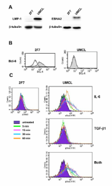
          Results confirmed the sensitivity of LCL8664, an immunoblastic SAIDS-NHL cell line, to TGF-beta1-mediated growth inhibition, and further demonstrated the partial rescue by simultaneous treatment with IL-6. IL-6 was shown to activate STAT3, even in the presence of TGF-beta1, and thereby to activate proliferative and anti-apoptotic pathways. By comparison, human AIDS-NHL cell lines differed in their responsiveness to TGF-beta1 and IL-6. Analysis of a recently derived AIDS-NHL cell line, UMCL01-101, indicated that it represents immunoblastic AIDS-DLCBL. Like LCL-8664, UMCL01-101 was sensitive to TGF-beta1-mediated inhibition, rescued partially by IL-6, and demonstrated rapid STAT3 activation following IL-6 treatment even in the presence of TGF-beta1.

          Conclusion

          These studies indicate that the sensitivity of immunoblastic AIDS- or SAIDS-DLBCL to TGF-beta1-mediated growth inhibition may be overcome through the stimulation of proliferative and anti-apoptotic signals by IL-6, particularly through the rapid activation of STAT3.

          Related collections

          Most cited references50

          • Record: found
          • Abstract: found
          • Article: not found

          Activated STAT signaling in human tumors provides novel molecular targets for therapeutic intervention.

          The signal transducers and activators of transcription (STAT)factors function as downstream effectors of cytokine and growth factor receptor signaling. Compared with normal cells and tissues, constitutively activated STATs have been detected in a wide variety of human cancer cell lines and primary tumors. STATs are activated by tyrosine phosphorylation, which is normally a transient and tightly regulated process. In tumor cells, constitutive activation of STATs is linked to persistent activity of tyrosine kinases, including Src, epidermal growth factor receptor, Janus kinases, Bcr-Abl, and many others. Such oncogenic tyrosine kinases are often activated as a consequence of permanent ligand/receptor engagement in autocrine or paracrine cytokine and growth factor signaling or represent autonomous constitutively active enzymes as a result of genetic alterations found in tumor but not normal cells. Persistent signaling of specific STATs, in particular Stat3 and Stat5, has been demonstrated to directly contribute to oncogenesis by stimulating cell proliferation and preventing apoptosis. STATs participate in oncogenesis through up-regulation of genes encoding apoptosis inhibitors and cell cycle regulators such as Bcl-x(L), Mcl-1, cyclins D1/D2, and c-Myc. Inhibition of constitutively active STAT signaling pathways has been shown repeatedly to inhibit tumor cell growth in vitro and in vivo and provides a novel means for therapeutic intervention in human cancer. In this review, we will: (a) explain the mechanisms of STAT activation in normal and malignant signaling; (b) summarize recent evidence for the critical role of constitutively activated Stat3 and Stat5 in oncogenesis; (c) identify candidate STAT target genes implicated in tumor progression; and (d) discuss molecular and pharmacological strategies to interfere with STAT signaling for potential therapeutic intervention in human cancer.
            Bookmark
            • Record: found
            • Abstract: found
            • Article: not found

            The role of STATs in transcriptional control and their impact on cellular function.

            The STAT proteins (Signal Transducers and Activators of Transcription), were identified in the last decade as transcription factors which were critical in mediating virtually all cytokine driven signaling. These proteins are latent in the cytoplasm and become activated through tyrosine phosphorylation which typically occurs through cytokine receptor associated kinases (JAKs) or growth factor receptor tyrosine kinases. Recently a number of non-receptor tyrosine kinases (for example src and abl) have been found to cause STAT phosphorylation. Phosphorylated STATs form homo- or hetero-dimers, enter the nucleus and working coordinately with other transcriptional co-activators or transcription factors lead to increased transcriptional initiation. In normal cells and in animals, ligand dependent activation of the STATs is a transient process, lasting for several minutes to several hours. In contrast, in many cancerous cell lines and tumors, where growth factor dysregulation is frequently at the heart of cellular transformation, the STAT proteins (in particular Stats 1, 3 and 5) are persistently tyrosine phosphorylated or activated. The importance of STAT activation to growth control in experiments using anti-sense molecules or dominant negative STAT protein encoding constructs performed in cell lines or studies in animals lacking specific STATs strongly indicate that STATs play an important role in controlling cell cycle progression and apoptosis. Stat1 plays an important role in growth arrest, in promoting apoptosis and is implicated as a tumor suppressor; while Stats 3 and 5 are involved in promoting cell cycle progression and cellular transformation and preventing apoptosis. Many questions remain including: (1) a better understanding of how the STAT proteins through association with other factors increase transcription initiation; (2) a more complete definition of the sets of genes which are activated by different STATs and (3) how these sets of activated genes differ as a function of cell type. Finally, in the context of many cancers, where STATs are frequently persistently activated, an understanding of the mechanisms leading to their constitutive activation and defining the potential importance of persistent STAT activation in human tumorigenesis remains. Oncogene (2000).
              Bookmark
              • Record: found
              • Abstract: found
              • Article: not found

              Constitutive activation of Stat3 signaling confers resistance to apoptosis in human U266 myeloma cells.

              Interleukin 6 (IL-6) is the major survival factor for myeloma tumor cells and induces signaling through the STAT proteins. We report that one STAT family member, Stat3, is constitutively activated in bone marrow mononuclear cells from patients with multiple myeloma and in the IL-6-dependent human myeloma cell line U266. Moreover, U266 cells are inherently resistant to Fas-mediated apoptosis and express high levels of the antiapoptotic protein Bcl-xL. Blocking IL-6 receptor signaling from Janus kinases to the Stat3 protein inhibits Bcl-xL expression and induces apoptosis, demonstrating that Stat3 signaling is essential for the survival of myeloma tumor cells. These findings provide evidence that constitutively activated Stat3 signaling contributes to the pathogenesis of multiple myeloma by preventing apoptosis.
                Bookmark

                Author and article information

                Journal
                BMC Cancer
                BMC Cancer
                BioMed Central (London )
                1471-2407
                2007
                26 February 2007
                : 7
                : 35
                Affiliations
                [1 ]Department of Microbiology & Immunology and Tulane Cancer Center, Tulane University Health Sciences Center, New Orleans, Louisiana, USA
                Article
                1471-2407-7-35
                10.1186/1471-2407-7-35
                1810304
                17324269
                d095f207-490f-4d7a-afcf-16c44d752019
                Copyright © 2007 Ruff et al; licensee BioMed Central Ltd.

                This is an Open Access article distributed under the terms of the Creative Commons Attribution License ( http://creativecommons.org/licenses/by/2.0), which permits unrestricted use, distribution, and reproduction in any medium, provided the original work is properly cited.

                History
                : 8 November 2006
                : 26 February 2007
                Categories
                Research Article

                Oncology & Radiotherapy
                Oncology & Radiotherapy

                Comments

                Comment on this article