- Record: found
- Abstract: found
- Article: found
Reinforcement of peritoneal repair in donor site post-concurrent laparotomy and rectus abdominis myocutaneous flap breast reconstruction using autologous dermal graft repair from zone 4 of deep inferior epigastric perforator flap: A case series in Asian patients
letter
Read this article at
There is no author summary for this article yet. Authors can add summaries to their articles on ScienceOpen to make them more accessible to a non-specialist audience.
Abstract
Sir,
With the rising incidence of breast cancer, the deep inferior epigastric perforator
(DIEP) flaps have become a popular autologous tissue transfer option for breast reconstruction
post-mastectomy. With the advent of prolene mesh reinforcement of abdominal wall repair
techniques, donor site bulging and herniation have largely been eliminated; however,
in specific clinical cases where there is weakening of the abdominal wall secondary
to previous abdominal surgery or presence of tumours, which require excision, these
complications may once again present.[1
2] Several techniques have been reported to reduce abdominal donor site morbidity,
post-DIEP flap harvesting for breast reconstruction, including placement of synthetic
or biologic meshes such as acellular dermal matrices.[3
4
5]
We illustrate our reconstructive approach in three patients, highlighting a novel
use of the dermal graft from zone 4 of the DIEP flap to reinforce abdominal wall repair
and to act as an adjunct to the mesh repair. In two of these cases, the primary DIEP
surgery was combined with a laparotomy, which is a complex situation. This is because
any dehiscence of the peritoneal repair will result in gut spillage into the abdominal
subcutaneous pocket. Moreover, the DIEP flap harvesting in itself causes a further
skin shortage which may cause peritoneal wound breakdowns. Dehiscence of the abdominal
wound would lead to an open abdomen and herniation of the bowel contents that would
be catastrophic.
A 47-year-old woman presented with right-sided breast cancer. A pre-operative computed-tomography
(CT) scan demonstrated an incidental urachal cyst. She was noted to have a significant
medical history of laparoscopic oophorectomy and a previous caesarean section. In
view of the malignant potential of her urachal cyst, it was planned for excision in
the same setting as the flap. A DIEP flap was performed and zone 4 was planned to
be discarded. Following the excision of the urachal cyst via a midline incision, a
peritoneal defect remained which was repaired primarily posteriorly and reinforced
with a dermal graft sublay technique, harvested from zone 4 of the flap, anchored
with prolene 2/0. The dermal graft was positioned with dermis facing up and fat facing
down towards the intraabdominal contents to decrease risk of adhesions. This is an
autologous repair technique which can be performed in the same sitting without additional
costs, unlike using a foreign mesh material such as Omyra[6] or double-layered non-adhesive
prolene meshes.
A 58-year-old woman presented with right-sided breast cancer and underwent a mastectomy
with DIEP flap reconstruction. During the process of raising the flap, inadvertent
breach of the peritoneal wall occurred due to adhesions that had formed secondary
to a caesarean section which patient had previously undergone. The risk of such a
breach is that bowel may herniate laterally and get trapped between prolene mesh if
it is used for repair and posterior sheath and cause bowel ischemia. Hence, the peritoneal
defect was repaired primarily and overlay technique reinforcement with dermal graft
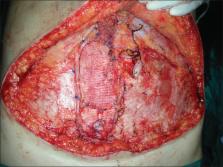
harvested from zone 4 of the flap was performed [Figures 1 and 2].
Figure 1
Dermal graft inset with prolene mesh overlying
Figure 2
Dermal graft and prolene mesh inset
A 41-year-old woman presented with left breast cancer post-mastectomy for DIEP reconstruction.
She had a significant history of hormonal therapy for the breast cancer, a left 5
cm endometriotic cyst and raising cancer antigen 125 trends, hence was also planned
for total abdominal hysterectomy and bilateral salpingo-oophrectomy in the same seating.
Following the raising of the DIEP flap, a paramedian incision was performed to access
the uterus and ovaries. In view of the large size of the peritoneal defect, a combination
reinforcement technique was adopted in this case. The dermal graft harvested from
zone 4 of the DIEP flap was laid over the inferior peritoneal incision and secured
with prolene 2/0. A prolene mesh was then laid over the peritoneal repair and the
rectus sheath was double breasted [Figures 3 and 4].
Figure 3
Double-breasting of the fascia
Figure 4
Computed tomography post-repair with dermal graft and prolene mesh inset
Therefore, we report three cases of peritoneal defect repair prolene reinforcement
with autologous dermal graft [Figures 1–3]. The dermal graft serves to reinforce the
primary peritoneal repair, which may be flimsy and prone to tearing and in the event
of peritoneal dehiscence allows the gut to be contained by the dermal graft. This
prevents direct contact of the gut with the overlying mesh. It allows for subsequent
repair and closure of the abdomen with negative pressure dressing therapy as the bowel
is contained. It avoids the complications associated with only repairing the sheath
with a mesh risking bowel being abraded by mesh used for fascial repair if it herniates
through weakened peritoneal lining or secondary peritoneal defect. The autologous
dermal graft onlay technique reduces the risk of post-operative ventral hernias by
reinforcing primary peritoneal repair. Other options are acellular dermal matrices
and alloplastic procide. Our autologous dermal graft is a form of matrix, which has
many potential uses yet to be explored. It is harvested from tissue that would otherwise
be discarded in the DIEP flap and is a safe option, which incurs no additional costs
to the patient and provides a heretofore untold benefit to the patient. In our series
of three patients, no abdominal wall weakness in the flap donor site was identified
following a series of clinical examinations for at least 12 months after autologous
dermal graft reinforcement of peritoneal wall defect. A well-formed and thickened
fascial layer at the abdominal donor fascial repair site was revealed on follow-up
by CT scans [Figure 4]. This objective finding, along with our clinical observation,
supports the use of dermal graft for repair of the abdominal donor site peritoneal
defect following flap harvesting. In conclusion, our two-layered technique of repair
prevents abdominal complications and is safe, by preventing direct contact of the
mesh with intra-abdominal contents and is cost effective as we are using a segment
of the DIEP flap which would otherwise usually be discarded.
Financial support and sponsorship
Nil.
Conflicts of interest
There are no conflicts of interest.
Related collections
Most cited references5
- Record: found
- Abstract: found
- Article: not found
Breast Reconstruction with the free TRAM or DIEP flap: patient selection, choice of flap, and outcome.
Maurice Nahabedian, Gregory Galdino, Bahram Momen … (2002)
- Record: found
- Abstract: found
- Article: not found
Abdominal wall strength, bulging, and hernia after TRAM flap breast reconstruction.
Roger G. Evans, Fernanda Kroll, G. Robb … (1995)
- Record: found
- Abstract: found
- Article: not found
Hernia prevention and aesthetic contouring of the abdomen following TRAM flap breast reconstruction by the use of polypropylene mesh.
R Zienowicz, Patrick J C May (1995)