2
views
0
recommends
+1 Recommend
0 collections
    0
    shares
      • Record: found
      • Abstract: found
      • Article: found
      Is Open Access

      New low-cost magnifying device for temporal bone laboratory

      research-article

      Read this article at

      Bookmark
          There is no author summary for this article yet. Authors can add summaries to their articles on ScienceOpen to make them more accessible to a non-specialist audience.

          Abstract

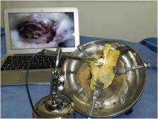
          Temporal bone dissection has important role in educating and training oto and skull base surgeons. Mounting of a temporal bone laboratory is expensive. A dedicated magnifying system, such as a surgical microscope or an endoscopic equipment, represents one of the most significant costs. The aim of this study is to test and demonstrate the utility of a commercial USB as a low-cost solution to equip the laboratory with a good magnifying system and illumination.

          Related collections

          Most cited references4

          • Record: found
          • Abstract: not found
          • Article: not found

          Resident Participation in Cadaveric Temporal Bone Dissection Correlates With Improved Performance on a Standardized Skill Assessment Instrument

            Bookmark
            • Record: found
            • Abstract: found
            • Article: found

            Cadaveric Temporal Bone Dissection: Is It Obsolete Today?

            Introduction Traditionally, surgical training in otology, is imparted by dissecting harvested human cadaveric temporal bones. However, maintenance of a cadaveric temporal bone laboratory is expensive and carries risk of exposure to infection. In recent times, other modalities of training are gaining ground and are likely to eventually replace cadaveric temporal bone dissection altogether. Objectives Other alternative methods of training are emerging. New technology like simulation and virtual reality as high-fidelity, safer alternatives, are making rapid strides as teaching tools. Other options are the use of animal temporal bones as teaching tools. The advantages of these are compared. Data Synthesis None of these modalities can replicate the innumerable anatomical variations which are a characteristic feature of the human temporal bone. A novice surgeon not only needs exposure to surgical anatomy and it's variations but also needs to develop hand-eye coordination skills to gain expertise. Conclusion Deliberate practice on human cadaveric temporal bones only, will confer both mastery in anatomy and surgical technique. The human cadaveric temporal bone is ideal simulator for training in otology.
              Bookmark
              • Record: found
              • Abstract: found
              • Article: not found

              Preparation of a temporal bone exhibit.

              Temporal bone dissection forms an important aspect in the training of an otolaryngologist. The more dissection one does the more confident one is in the operating room. The aim of this paper is to advise in the preparation of temporal bones for the purpose of display, exhibition or competition. The practical aspects of dissection are deliberately avoided concentrating on the selection of bones pre- and post-dissection, preparation of the bones, fixing and display of vessels and nerves and also mounting and lighting for exhibition purposes. The temporal bone laboratory should be well equipped with a microscope, a power drill with a range of cutting and polishing burrs, a range of fine instruments, a suction machine with different sizes of suction tips, water source, a place to store the bones etc., thus creating an ideal environment for temporal bone dissection.
                Bookmark

                Author and article information

                Contributors
                Journal
                J Otol
                J Otol
                Journal of Otology
                Chinese PLA General Hospital
                1672-2930
                23 February 2019
                June 2019
                23 February 2019
                : 14
                : 2
                : 73-75
                Affiliations
                [a ]Unit of Otolaryngology, UOS ORL TI, Campus Bio-Medico University, Rome, Italy
                [b ]Department of Otolaryngology, University of Foggia, Foggia, Italy
                [c ]ORL ROMA Care & Research, Rome, Italy
                [d ]Department of Otolaryngology, Department of Clinical Sciences and Community Health, Fondazione I.R.C.C.S. Ca’ Granda, Ospedale Maggiore Policlinico, University of Milan, Milan, Italy
                [e ]Institut G. Portmann, 114 avenue d'Arès, 33076, Bordeaux cedex, France
                Author notes
                []Corresponding author. Department of Otolaringology, University of Foggia, 71122, Foggia, Italy. moffa.antonio1@ 123456gmail.com
                Article
                S1672-2930(19)30015-7
                10.1016/j.joto.2019.02.001
                6570644
                d164f23a-9207-4cce-aa7c-d79a5a950320
                © 2019 PLA General Hospital Department of Otolaryngology Head and Neck Surgery. Production and hosting by Elsevier (Singapore) Pte Ltd.

                This is an open access article under the CC BY-NC-ND license (http://creativecommons.org/licenses/by-nc-nd/4.0/).

                History
                : 30 January 2019
                : 10 February 2019
                : 20 February 2019
                Categories
                Research Article

                temporal bone dissection,temporal bone laboratory,temporal bone surgical technique training

                Comments

                Comment on this article