4
views
0
recommends
+1 Recommend
0 collections
    0
    shares
      • Record: found
      • Abstract: found
      • Article: found
      Is Open Access

      Prediction of multivessel coronary artery disease and candidates for stress-only imaging using multivariable models with myocardial perfusion imaging

      research-article

      Read this article at

      Bookmark
          There is no author summary for this article yet. Authors can add summaries to their articles on ScienceOpen to make them more accessible to a non-specialist audience.

          Abstract

          Purpose

          Selecting patients with coronary multivessel disease (MVD) or no stenosis using myocardial perfusion imaging (MPI) is challenging. We aimed to create a model to predict MVD using a combination of quantitative MPI values and background factors of patients. We also assessed whether patients in the same database could be selected who do not require rest studies (stress-only imaging).

          Methods

          We analyzed data from 1001 patients who had been assessed by stress MPI at 12 centers and 463 patients who had not undergone revascularization in Japan. Quantitative values based on MPI were obtained using cardioREPO software, which included myocardial perfusion defect scores, left ventricular ejection fractions and volumes. Factors in MPI and clinical backgrounds that could predict MVD were investigated using univariate and multivariate analyses. We also investigated whether stress data alone could predict patients without coronary stenosis to identify candidates for stress-only imaging.

          Results

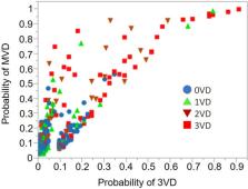
          We selected summed stress score (SSS), rest end-diastolic volume, and hypertension to create a predictive model for MVD. A logistic regression model was created with an area under the receiver operating characteristics curve (AUC) of 0.825. To more specifically predict coronary three-vessel disease, the AUC was 0.847 when SSS, diabetes, and hypertension were selected. The mean probabilities of abnormality based on the MVD prediction model were 12%, 24%, 40%, and 51% for no-, one-, two-, and three-vessel disease, respectively ( p < 0.0001). For the model to select patients with stress-only imaging, the AUC was 0.78 when the model was created using SSS, stress end-systolic volume and the number of risk factors (diabetes, hypertension, chronic kidney disease, and a history of smoking).

          Conclusion

          A model analysis combining myocardial SPECT and clinical data can predict MVD, and can select patients for stress-only tests. Our models should prove useful for clinical applications.

          Related collections

          Most cited references35

          • Record: found
          • Abstract: found
          • Article: not found

          Incremental prognostic value of myocardial perfusion single photon emission computed tomography for the prediction of cardiac death: differential stratification for risk of cardiac death and myocardial infarction.

          The incremental prognostic value of stress single photon emission computed tomography (SPECT) for the prediction of cardiac death as an individual end point and the implications for risk stratification are undefined. We identified 5183 consecutive patients who underwent stress/rest SPECT and were followed up for the occurrence of cardiac death or myocardial infarction. Over a mean follow up of 642+/-226 days, 119 cardiac deaths and 158 myocardial infarctions occurred (3.0% cardiac death rate, 2.3% myocardial infarction rate). Patients with normal scans were at low risk (< or =0.5%/y), and rates of both outcomes increased significantly with worsening scan abnormalities. Patients who underwent exercise stress and had mildly abnormal scans had low rates of cardiac death but higher rates of myocardial infarction (0.7%/y versus 2.6%/y; P<.05). After adjustment for prescan information, scan results provided incremental prognostic value toward the prediction of cardiac death. The identification of patients at intermediate risk of nonfatal myocardial infarction and low risk for cardiac death by SPECT may result in significant cost savings when applied to a clinical testing strategy. Myocardial perfusion SPECT yields incremental prognostic information toward the identification of cardiac death. Patients with mildly abnormal scans after exercise stress are at low risk for cardiac death but intermediate risk for nonfatal myocardial infarction and thus may benefit from a noninvasive strategy and may not require invasive management.
            Bookmark
            • Record: found
            • Abstract: not found
            • Article: not found

            Single Photon Emission Computed Tomography (SPECT) Myocardial Perfusion Imaging Guidelines: Instrumentation, Acquisition, Processing, and Interpretation

              Bookmark
              • Record: found
              • Abstract: found
              • Article: found
              Is Open Access

              High-Dose Versus Low-Dose Pitavastatin in Japanese Patients With Stable Coronary Artery Disease (REAL-CAD)

              Supplemental Digital Content is available in the text.
                Bookmark

                Author and article information

                Contributors
                kunita.nmd@gmail.com
                nakajima@med.kanazawa-u.ac.jp
                Journal
                Ann Nucl Med
                Ann Nucl Med
                Annals of Nuclear Medicine
                Springer Nature Singapore (Singapore )
                0914-7187
                1864-6433
                5 June 2022
                5 June 2022
                2022
                : 36
                : 7
                : 674-683
                Affiliations
                [1 ]GRID grid.412002.5, ISNI 0000 0004 0615 9100, Department of Nuclear Medicine, , Kanazawa University Hospital, ; Kanazawa, Japan
                [2 ]GRID grid.9707.9, ISNI 0000 0001 2308 3329, Department of Functional Imaging and Artificial Intelligence, , Kanazawa University, ; Kanazawa, Japan
                [3 ]Department of Cardiovascular Medicine, Hakodate Goryoukaku Hospital, Hakodate, Japan
                [4 ]GRID grid.174567.6, ISNI 0000 0000 8902 2273, Department of Radioisotope Medicine, Atomic Bomb Disease and Hibakusha Medicine Unit, Atomic Bomb Disease Institute, , Nagasaki University, ; Nagasaki, Japan
                Author information
                http://orcid.org/0000-0002-4802-7857
                Article
                1751
                10.1007/s12149-022-01751-7
                9226096
                35661104
                d1a6fdbb-ac8f-413e-954b-e773dd3b75fa
                © The Author(s) 2022

                Open AccessThis article is licensed under a Creative Commons Attribution 4.0 International License, which permits use, sharing, adaptation, distribution and reproduction in any medium or format, as long as you give appropriate credit to the original author(s) and the source, provide a link to the Creative Commons licence, and indicate if changes were made. The images or other third party material in this article are included in the article's Creative Commons licence, unless indicated otherwise in a credit line to the material. If material is not included in the article's Creative Commons licence and your intended use is not permitted by statutory regulation or exceeds the permitted use, you will need to obtain permission directly from the copyright holder. To view a copy of this licence, visit http://creativecommons.org/licenses/by/4.0/.

                History
                : 30 January 2022
                : 2 May 2022
                Funding
                Funded by: FundRef http://dx.doi.org/10.13039/501100012269, Science and Technology Innovative Research Team in Higher Educational Institutions of Hunan Province;
                Award ID: 20K07990
                Award Recipient :
                Categories
                Original Article
                Custom metadata
                © The Author(s) under exclusive licence to The Japanese Society of Nuclear Medicine 2022

                Radiology & Imaging
                coronary artery disease,single-photon emission computed tomography,multivariable model,quantitation

                Comments

                Comment on this article