0
views
0
recommends
+1 Recommend
0 collections
    0
    shares
      • Record: found
      • Abstract: found
      • Article: found
      Is Open Access

      Characteristics and Mechanism of Upper Airway Collapse Revealed by Dynamic MRI During Natural Sleep in Patients with Severe Obstructive Sleep Apnea

      research-article

      Read this article at

      Bookmark
          There is no author summary for this article yet. Authors can add summaries to their articles on ScienceOpen to make them more accessible to a non-specialist audience.

          Abstract

          Purpose

          Upper airway collapse during sleep in patients with obstructive sleep apnea (OSA) is a complex and dynamic phenomenon. By observing and analyzing the dynamic changes in the upper airway and its surrounding tissues during airway obstruction, we aim to reveal dynamic characteristics in different obstruction patterns, and the relationship between anatomical features during normal breathing and dynamic characteristics of airway obstruction.

          Patients and Methods

          Dynamic MRI was performed in 23 male patients (age range 26–63) with severe OSA diagnosed by overnight polysomnography, and obstruction events were identified from their images. Dynamic changes in parameters of the upper airway and surrounding tissues were measured to assess the key characteristics in different obstruction patterns.

          Results

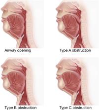
          We categorized airway obstruction into four types based on the obstruction location and motion characteristics of tissues during collapse, and detailed the alterations in the airway and surrounding tissues under each obstruction pattern. In all 112 obstruction events extracted from the dynamic images of 23 patients, type A (retropalatal obstruction caused by the soft palate separated from the tongue), BI, BII (both retropalatal obstructions caused by the soft palate attached to the tongue, and C (retropalatal and retroglossal obstruction caused by the soft palate and the tongue), accounted for 28.6%, 44.6%, 12.5%, and 14.3% respectively. In severe OSA patients with tongue and palatal obstruction related to type B or C, the more posterior hyoid position, shorter distance between tongue and uvula, and wider retropalatal space, the larger the tongue displacement and deformation during collapse, and the greater the reduction in airway space.

          Conclusion

          There are multiple airway obstruction patterns, each with its own anatomical characteristics and behaviors during collapse. Hyoid position, tongue and uvula distance, and retropalatal space play an important role in airway collapse and should be paid more attention in the treatment of OSA.

          Related collections

          Most cited references40

          • Record: found
          • Abstract: found
          • Article: not found

          Diagnosis and Management of Obstructive Sleep Apnea: A Review

          Obstructive sleep apnea (OSA) affects 17% of women and 34% of men in the US and has a similar prevalence in other countries. This review provides an update on the diagnosis and treatment of OSA.
            Bookmark
            • Record: found
            • Abstract: found
            • Article: not found

            Obesity and obstructive sleep apnea: pathogenic mechanisms and therapeutic approaches.

            Obstructive sleep apnea is a common disorder whose prevalence is linked to an epidemic of obesity in Western society. Sleep apnea is due to recurrent episodes of upper airway obstruction during sleep that are caused by elevations in upper airway collapsibility during sleep. Collapsibility can be increased by underlying anatomic alterations and/or disturbances in upper airway neuromuscular control, both of which play key roles in the pathogenesis of obstructive sleep apnea. Obesity and particularly central adiposity are potent risk factors for sleep apnea. They can increase pharyngeal collapsibility through mechanical effects on pharyngeal soft tissues and lung volume, and through central nervous system-acting signaling proteins (adipokines) that may affect airway neuromuscular control. Specific molecular signaling pathways encode differences in the distribution and metabolic activity of adipose tissue. These differences can produce alterations in the mechanical and neural control of upper airway collapsibility, which determine sleep apnea susceptibility. Although weight loss reduces upper airway collapsibility during sleep, it is not known whether its effects are mediated primarily by improvement in upper airway mechanical properties or neuromuscular control. A variety of behavioral, pharmacologic, and surgical approaches to weight loss may be of benefit to patients with sleep apnea, through distinct effects on the mass and activity of regional adipose stores. Examining responses to specific weight loss strategies will provide critical insight into mechanisms linking obesity and sleep apnea, and will help to elucidate the humoral and molecular predictors of weight loss responses.
              Bookmark
              • Record: found
              • Abstract: found
              • Article: not found

              Identification of upper airway anatomic risk factors for obstructive sleep apnea with volumetric magnetic resonance imaging.

              We used sophisticated volumetric analysis techniques with magnetic resonance imaging in a case-control design to study the upper airway soft tissue structures in 48 control subjects (apnea-hypopnea index, 2.0 +/- 1.6 events/hour) and 48 patients with sleep apnea (apnea-hypopnea index, 43.8 +/- 25.4 events/hour). Our design used exact matching on sex and ethnicity, frequency matching on age, and statistical control for craniofacial size and visceral neck fat. The data support our a priori hypotheses that the volume of the soft tissue structures surrounding the upper airway is enlarged in patients with sleep apnea and that this enlargement is a significant risk factor for sleep apnea. After covariate adjustments the volume of the lateral pharyngeal walls (p < 0.0001), tongue (p < 0.0001), and total soft tissue (p < 0.0001) was significantly larger in subjects with sleep apnea than in normal subjects. These data also demonstrated, after covariate adjustments, significantly increased risk of sleep apnea the larger the volume of the tongue, lateral pharyngeal walls, and total soft tissue: (1) total lateral pharyngeal wall (odds ratio [OR], 6.01; 95% confidence interval [CI], 2.62-17.14); (2) total tongue (OR, 4.66; 95% CI, 2.31-10.95); and (3) total soft tissue (OR, 6.95; 95% CI, 3.08-19.11). In a multivariable logistic regression analysis the volume of the tongue and lateral walls was shown to independently increase the risk of sleep apnea.
                Bookmark

                Author and article information

                Journal
                Nat Sci Sleep
                Nat Sci Sleep
                nss
                Nature and Science of Sleep
                Dove
                1179-1608
                01 November 2023
                2023
                : 15
                : 885-902
                Affiliations
                [1 ]School of Biomedical Engineering, Capital Medical University , Beijing, People’s Republic of China
                [2 ]Beijing Key Laboratory of Fundamental Research on Biomechanics in Clinical Application, Capital Medical University , Beijing, People’s Republic of China
                [3 ]Department of Radiology, Beijing Tongren Hospital, Capital Medical University , Beijing, People’s Republic of China
                Author notes
                Correspondence: Yaqi Huang, School of Biomedical Engineering, Capital Medical University , Beijing, People’s Republic of China, Tel +86-10-83911809, Email yqhuang@ccmu.edu.cn
                Junfang Xian, Department of Radiology, Beijing Tongren Hospital, Capital Medical University , Beijing, People’s Republic of China, Email cjr.xianjunfan@vip.163.com
                [*]

                These authors contributed equally to this work

                Author information
                http://orcid.org/0000-0002-3790-1502
                http://orcid.org/0009-0002-1357-1980
                http://orcid.org/0000-0002-8366-4245
                http://orcid.org/0000-0003-2191-9393
                http://orcid.org/0000-0001-6811-5021
                Article
                423303
                10.2147/NSS.S423303
                10625767
                d23d1afc-ea58-4243-9081-a4162dbfcbd1
                © 2023 Li et al.

                This work is published and licensed by Dove Medical Press Limited. The full terms of this license are available at https://www.dovepress.com/terms.php and incorporate the Creative Commons Attribution – Non Commercial (unported, v3.0) License ( http://creativecommons.org/licenses/by-nc/3.0/). By accessing the work you hereby accept the Terms. Non-commercial uses of the work are permitted without any further permission from Dove Medical Press Limited, provided the work is properly attributed. For permission for commercial use of this work, please see paragraphs 4.2 and 5 of our Terms ( https://www.dovepress.com/terms.php).

                History
                : 28 May 2023
                : 19 October 2023
                Page count
                Figures: 10, Tables: 2, References: 40, Pages: 18
                Funding
                Funded by: supported by the National Natural Science Foundation of China;
                This work was supported by the National Natural Science Foundation of China (Grant Numbers: 34670959, 81171422, 11902209).
                Categories
                Original Research

                airway obstruction patterns,obstruction location,tissue motion characteristics,hyoid position,retropalatal space

                Comments

                Comment on this article