7
views
0
recommends
+1 Recommend
0 collections
    0
    shares
      • Record: found
      • Abstract: found
      • Article: found
      Is Open Access

      Analysis of acetabulum in children with developmental dysplasia of the hip by MRI scan

      research-article

      Read this article at

      Bookmark
          There is no author summary for this article yet. Authors can add summaries to their articles on ScienceOpen to make them more accessible to a non-specialist audience.

          Abstract

          To review the value of acetabular magnetic resonance imaging (MRI) in children with developmental dysplasia of the hip (DDH) of different ages.

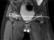
          Eighty-eight medical records of children with unilateral DDH who were diagnosed and treated in our hospital between January 2010 and December 2015 were retrospectively analyzed. The affected hips were put into the case group, and the normal hips were put into the control group. All cases were further divided into 3 age groups: infant (<1 year), 16 cases; young children (1–3 years), 48 cases; and children (3–13 years), 24 cases. The differences of the acetabular depth (AD), the bony acetabular index (BAI), and the cartilaginous acetabular index (CAI) between each group were measured and compared for a linear correlation analysis. At the same time, the distribution of the acetabular cartilage in the anterosuperior, top, and posterosuperior parts (the three parts) from the two groups was measured, respectively.

          Measurement results from both the case and control groups were as follows: AD was 5.46 ± 2.62 mm and 9.74 ± 2.33 mm; BAI was 33.26 ± 5.49° and 23.50 ± 5.33°; and CAI was 21.04 ± 6.16° and 12.71 ± 4.83°. Differences from the two groups were statistically significant ( t = 11.94, 13.78, 9.16, P < .05); BAI and CAI were linearly correlated ( r = 0.86, 0.75, P < .05). The AD in infant, young children, and children groups from the case group were 4.26 ± 0.42 mm, 4.79 ± 1.74 mm, and 7.31 ± 2.74 mm, respectively, which was statically significant as well ( F = 11.37, P < .05). Under the same grouping criteria, BAI was recorded as 29.04 ± 5.11°, 34.56 ± 4.27°, and 33.12 ± 5.69°; CAI was recorded as 16.62 ± 5.50°, 21.79 ± 6.33°, and 20.91 ± 6.40° separately. There was a linear correlation ( r = 0.78, 0.65, P < .05) between BAI and CAI in young children and children groups. The distribution of acetabular cartilage in the above-mentioned three parts from both young children and children groups was statistically significant ( P < .05).

          MRI is a satisfactory imaging modality to children with DDH of different ages for the assessment of AD, BAI, CAI, and acetabular cartilage in multiple locations. It can provide ample imaging reference to clinical evaluation of the acetabulum development in DDH.

          Related collections

          Most cited references19

          • Record: found
          • Abstract: found
          • Article: not found

          Imaging evaluation of developmental hip dysplasia in the young adult.

          The purpose of this article is to review the clinical and imaging features as well as the potential complications of hip dysplasia in the young adult. Hip dysplasia is an important cause of secondary osteoarthrosis, which accounts for a significant proportion of patients requiring total hip arthroplasty. The radiographic diagnosis of mild hip dysplasia in the young adult may be subtle and is primarily based on the detection of deficient coverage of the femoral head by the acetabulum. Cross-sectional imaging, including CT and MRI, afford improved detection and characterization by providing morphologic information about acetabular deficiency. MRI also allows evaluation of potential associated injuries to the articular cartilage, the labrum, and the ligamentum teres. Familiarity with the radiographic and cross-sectional imaging findings of mild hip dysplasia in the young adult may allow a timely diagnosis and implementation of treatment strategies, which may prevent or delay the development of early osteoarthritis.
            Bookmark
            • Record: found
            • Abstract: found
            • Article: not found

            Patterns of joint damage seen on MRI in early hip osteoarthritis due to structural hip deformities.

            The aim of this study was to evaluate differences in damage patterns assessed using magnetic resonance imaging (MRI) between hips with femoroacetabular impingement (FAI) and developmental dysplasia of the hip (DDH) as well as to correlate MRI findings with delayed Gadolinium enhanced MRI of cartilage (dGEMRIC) and with patient pain. This retrospective study included 40 patients (mean age 28.6 ± 11.2 years) who underwent dGEMRIC and morphological MRI of the hip. Twenty-one hips with FAI and 19 with DDH were investigated. A self-developed morphological grading (MRI score) and dGEMRIC evaluation were done on seven radial reformats obtained from an isotropic 3D True-fast imaging with steady state precession (FISP) sequence and an isotropic T1-mapping sequence. The observed damage patterns were summed up into sub-scores and a total MRI score. Labrum damage, paralabral cysts, and acetabular rim bone cysts were more common in DDH patients than in FAI patients. No significant differences were seen in the occurrence of cartilage damage, bone cysts, or osteophytes. In DDH (but not in FAI), the dGEMRIC index demonstrated a tendency for lower values in areas next to cartilage defects. There was no association between labrum damage and dGEMRIC index. A moderate correlation was seen between Western Ontario and McMaster Universities (WOMAC) pain score and cartilage damage, paralabral cysts, and the total MRI score. This study confirms a higher prevalence of labrum damage but not cartilage damage in patients with DDH in comparison to patients with FAI. In addition, our data suggests an association of cartilage damage and paralabral cysts with patient reported pain. Copyright © 2012 Osteoarthritis Research Society International. Published by Elsevier Ltd. All rights reserved.
              Bookmark
              • Record: found
              • Abstract: found
              • Article: not found

              Development of the osseous and cartilaginous acetabular index in normal children and those with developmental dysplasia of the hip: a cross-sectional study using MRI.

              The purpose of this study was to investigate the development of the osseous acetabular index (OAI) and cartilaginous acetabular index (CAI) using MRI. The OAI and CAI were measured on the coronal MR images of the hip in 81 children with developmental dysplasia of the hip (DDH), with a mean age of 19.6 months (3 to 70), and 241 normal control children with a mean age of 5.1 years (1 month to 12.5 years). Additionally the developmental patterns of the OAI and CAI in normal children were determined by age-based cross-sectional analysis. Unlike the OAI, the normal CAI decreased rapidly from a mean of 10.17° (sd 1.60) to a mean of 8.25° (sd 1.90) within the first two years of life, and then remained constant at a mean of 8.04° (sd 1.65) until adolescence. Although no difference in OAI was found between the uninvolved hips in children with unilateral DDH and normal hips (p = 0.639), the CAI was significantly different between them both (p < 0.001). The normal CAI has fully formed at birth, and is maintained constantly throughout childhood. The CAI in the unaffected hips in children with unilateral DDH is also mildly dysplastic.
                Bookmark

                Author and article information

                Journal
                Medicine (Baltimore)
                Medicine (Baltimore)
                MEDI
                Medicine
                Wolters Kluwer Health
                0025-7974
                1536-5964
                January 2019
                18 January 2019
                : 98
                : 3
                : e14054
                Affiliations
                Children's Hospital of Nanjing Medical University, Nanjing, Jiangsu Province, China.
                Author notes
                []Correspondence: Bo Wang, Department of Orthopedic, Children's Hospital of Nanjing Medical University, 72 Guangzhou Road, Nanjing 210008, Jiangsu Province, China (e-mail: bowangseu@ 123456163.com ).
                Article
                MD-D-17-04778 14054
                10.1097/MD.0000000000014054
                6370164
                30653114
                d4ebaf57-b4f9-423f-b8c9-66a4bca26fd2
                Copyright © 2019 the Author(s). Published by Wolters Kluwer Health, Inc.

                This is an open access article distributed under the terms of the Creative Commons Attribution-Non Commercial-No Derivatives License 4.0 (CCBY-NC-ND), where it is permissible to download and share the work provided it is properly cited. The work cannot be changed in any way or used commercially without permission from the journal. http://creativecommons.org/licenses/by-nc-nd/4.0

                History
                : 1 August 2017
                : 7 December 2018
                : 17 December 2018
                Categories
                6200
                Research Article
                Observational Study
                Custom metadata
                TRUE

                acetabulum,children,developmental dysplasia of the hip (ddh),magnetic resonance imaging (mri)

                Comments

                Comment on this article