15
views
0
recommends
+1 Recommend
0 collections
    0
    shares
      • Record: found
      • Abstract: found
      • Article: found
      Is Open Access

      Improved Software to Browse the Serial Medical Images for Learning

      research-article

      Read this article at

      Bookmark
          There is no author summary for this article yet. Authors can add summaries to their articles on ScienceOpen to make them more accessible to a non-specialist audience.

          Abstract

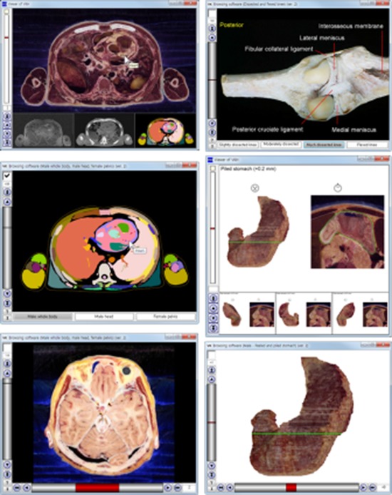
          The thousands of serial images used for medical pedagogy cannot be included in a printed book; they also cannot be efficiently handled by ordinary image viewer software. The purpose of this study was to provide browsing software to grasp serial medical images efficiently. The primary function of the newly programmed software was to select images using 3 types of interfaces: buttons or a horizontal scroll bar, a vertical scroll bar, and a checkbox. The secondary function was to show the names of the structures that had been outlined on the images. To confirm the functions of the software, 3 different types of image data of cadavers (sectioned and outlined images, volume models of the stomach, and photos of the dissected knees) were inputted. The browsing software was downloadable for free from the homepage (anatomy.co.kr) and available off-line. The data sets provided could be replaced by any developers for their educational achievements. We anticipate that the software will contribute to medical education by allowing users to browse a variety of images.

          Graphical Abstract

          Related collections

          Most cited references15

          • Record: found
          • Abstract: found
          • Article: not found

          Stereotaxic display of brain lesions.

          Traditionally lesion location has been reported using standard templates, text based descriptions or representative raw slices from the patient's CT or MRI scan. Each of these methods has drawbacks for the display of neuroanatomical data. One solution is to display MRI scans in the same stereotaxic space popular with researchers working in functional neuroimaging. Presenting brains in this format is useful as the slices correspond to the standard anatomical atlases used by neuroimagers. In addition, lesion position and volume are directly comparable across patients. This article describes freely available software for presenting stereotaxically aligned patient scans. This article focuses on MRI scans, but many of these tools are also applicable to other modalities (e.g. CT, PET and SPECT). We suggest that this technique of presenting lesions in terms of images normalized to standard stereotaxic space should become the standard for neuropsychological studies.
            Bookmark
            • Record: found
            • Abstract: found
            • Article: not found

            Visible Korean human: improved serially sectioned images of the entire body.

            The data from the Visible Human Project (VHP) and the Chinese Visible Human (CVH), which are the serially sectioned images of the entire cadaver, are being used to produce three-dimensional (3-D) images and software. The purpose of our research, the Visible Korean Human (VKH), is to produce an enhanced version of the serially sectioned images of an entire cadaver that can be used to upgrade the 3-D images and software. These improvements are achieved without drastically changing the methods developed for the VHP and CVH; thus, a complementary solution was found. A Korean male cadaver was chosen without anything perfused into the cadaver; the entire body was magnetic resonance (MR) and computed tomography (CT) scanned at 1.0-mm intervals to produce MR and CT images. After scanning, entire body of the cadaver was embedded and serially sectioned at 0.2-mm intervals; each sectioned surface was inputted into a personal computer to produce anatomical images (pixel size: 0.2 mm) without any missing images. Eleven anatomical organs in the anatomical images were segmented to produce segmented images. The anatomical and segmented images were stacked and reconstructed to produce 3-D images. The VKH is an ongoing research; we will produce a female version of the VKH and provide more detailed segmented images. The data from the VHP, CVH, and VKH will provide valuable resources to the medical image library of 3-D images and software in the field of medical education and clinical trials.
              Bookmark
              • Record: found
              • Abstract: found
              • Article: not found

              Teaching anatomy: cadavers vs. computers?

              Our study was aimed to show if cadaver dissections are still important in the Anatomy Course for medical students or whether computerized resources could replace them. We followed three groups, one of them (698 students) proceeded through the Anatomy Course in a traditional way, meaning, with cadaver material enough to observe all the regions and structures; the second group (330 students) used many technological resources but not cadaver dissections; and the third group (145 students) followed the course, recently, with the same program but with both practical resources. Theoretical contents were developed in the same way and by the same professor. The traditional teaching group obtained better results than the technologically supported group, evaluated by the number of students that passed their exams. The third group results were better than the others, with regard to passed exams and marks. Even when computerized improvements have developed a new area giving students a lot of elements to facilitate their approach to imaging structures, the possibility of direct contact with tissues and anatomical elements cannot yet be replaced. We are demonstrating that the best possibility is the correct association of all these resources to complement one another.
                Bookmark

                Author and article information

                Journal
                J Korean Med Sci
                J. Korean Med. Sci
                JKMS
                Journal of Korean Medical Science
                The Korean Academy of Medical Sciences
                1011-8934
                1598-6357
                July 2017
                26 May 2017
                : 32
                : 7
                : 1195-1201
                Affiliations
                [1 ]Department of Computer Engineering, Inha University, Incheon, Korea.
                [2 ]Department of Anatomy, Ajou University School of Medicine, Suwon, Korea.
                [3 ]Department of Anatomy, Dongguk University School of Medicine, Gyeongju, Korea.
                Author notes
                Address for Correspondence: Beom Sun Chung, MD. Department of Anatomy, Ajou University School of Medicine, 164 World cup-ro, Yeongtong-gu, Suwon 16499, Republic of Korea. bschung@ 123456ajou.ac.kr
                Author information
                https://orcid.org/0000-0002-2467-5809
                https://orcid.org/0000-0002-0527-9763
                https://orcid.org/0000-0001-7956-4148
                https://orcid.org/0000-0001-7742-4846
                https://orcid.org/0000-0002-3644-9120
                Article
                10.3346/jkms.2017.32.7.1195
                5461326
                28581279
                d5aed1ef-094f-4e76-bb49-440947fa7ff0
                © 2017 The Korean Academy of Medical Sciences.

                This is an Open Access article distributed under the terms of the Creative Commons Attribution Non-Commercial License ( https://creativecommons.org/licenses/by-nc/4.0/) which permits unrestricted non-commercial use, distribution, and reproduction in any medium, provided the original work is properly cited.

                History
                : 07 February 2017
                : 16 April 2017
                Funding
                Funded by: National Research Foundation of Korea, CrossRef http://dx.doi.org/10.13039/501100003725;
                Award ID: 2015R1A2A2A01008248
                Funded by: Ministry of Trade, Industry and Energy, CrossRef http://dx.doi.org/10.13039/501100003052;
                Award ID: N0002249
                Funded by: Korea Institute for Advancement of Technology, CrossRef http://dx.doi.org/10.13039/501100003661;
                Award ID: N0002249
                Categories
                Original Article
                Medical Imaging

                Medicine
                visible human projects,cadaver,anatomy,body image,computer software
                Medicine
                visible human projects, cadaver, anatomy, body image, computer software

                Comments

                Comment on this article