2
views
0
recommends
+1 Recommend
0 collections
    0
    shares
      • Record: found
      • Abstract: found
      • Article: found
      Is Open Access

      Fenestration of the posterior cerebral artery P1-P2 junction: A case report

      case-report

      Read this article at

      Bookmark
          There is no author summary for this article yet. Authors can add summaries to their articles on ScienceOpen to make them more accessible to a non-specialist audience.

          Abstract

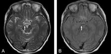
          Fenestration is a rare anatomical variation in the posterior cerebral artery. To the best of our knowledge, there are few reports of fenestrations at the posterior cerebral artery P1-P2 junction. Herein, we report a case of fenestration of the posterior cerebral artery P1-P2 junction diagnosed by 3-T magnetic resonance imaging and magnetic resonance angiography. A 75-year-old woman visited our hospital because of headaches. Magnetic resonance imaging incidentally showed fenestration around the P1-P2 segment of the right posterior cerebral artery. Magnetic resonance angiography revealed a small fenestration at the right posterior cerebral artery P1-P2 junction. The vessel diameter of both limbs forming the fenestration was nearly equal. Careful imaging assessment is important to identify fenestration of the posterior cerebral artery P1-P2 junction. Both magnetic resonance angiography and magnetic resonance imaging were useful for diagnosis in this case.

          Related collections

          Most cited references9

          • Record: found
          • Abstract: found
          • Article: not found

          Microsurgical anatomy of the upper basilar artery and the posterior circle of Willis.

          The microvascular anatomy of the posterior part of the circle of Willis, important in surgery of pituitary tumors and basilar aneurysms, was defined in 50 cadaver brains. Significant findings were as follows: 1) Anomalies of the posterior half of the circle of Willis were found in 46% of cases. 2) Hypoplastic P-1 (posterior cerebral segment) and posterior communicating segments gave origin to the same number and size of perforating arteries, having the same termination as normal-sized segments. Thus hypoplastic segments should be handled with care and divided to aid in exposure of the basilar bifurcation only after careful consideration. 3) An average of four perforating branches arose from P-1; most from the superior and posterior sufaces. No branches arose from the anterior surface of the basilar bifurcation. The most proximal P-1 branch originated 2 to 3 mm distal to the basilar bifurcation. It was most commonly a thalamoperforating artery. The largest P-1 branch was usually a thalamoperforating or a posterior choroidal artery. 4) An average of seven branches emerged from the superior and lateral surfaces of the posterior communicating artery. The anterior half was a richer source of perforators than the posterior half. The largest communicating branch in 80% of specimens supplied the premamillary area. 5) The anterior choroidal artery originated from the carotid artery on both sides in all cases. A double anterior choroidal artery was present in 4% of cases.
            Bookmark
            • Record: found
            • Abstract: found
            • Article: not found

            Cerebral arterial fenestrations.

            Arterial fenestrations are an anatomic variant with indeterminate significance. Given the controversy surrounding fenestrations we sought their prevalence within our practice along with their association with other cerebrovascular anomalies. We retrospectively reviewed 10,927 patients undergoing digital subtraction angiography between 1992 and 2011. Dictated reports were searched for the terms "fenestration" or "fenestrated" with images reviewed for relevance, yielding 228 unique cases. A Medline database search from February 1964 to January 2013 generated 304 citations, 127 cases of which were selected for analysis. Cerebral arterial fenestrations were identified in 228 patients (2.1%). At least one aneurysm was noted in 60.5% of patients, with an aneurysm arising from the fenestration in 19.6% of patients. Aneurysmal subarachnoid hemorrhage or non-aneurysmal subarachnoid hemorrhage were present in 60.1% and 15.8%, respectively. For the subset of patients with an aneurysm arising directly from a fenestration relative to those patients with an aneurysm not immediately associated with a fenestration, the prevalence of aneurysmal subarachnoid hemorrhage was 66.7% vs. 58.6% (p = 0.58). Fenestrations were more often within the posterior circulation (73.2%) than the anterior circulation (24.6%), though there was no difference in the prevalence of aneurysms within these groups (61.1% vs. 60.7%, p = 1.0). Cerebral arterial fenestrations are an anatomic variant more often manifesting at the anterior communicating arterial complex and basilar artery and with no definite pathological relationship with aneurysms.
              Bookmark
              • Record: found
              • Abstract: found
              • Article: not found

              Anomalies of the P1 segment of the posterior cerebral artery: early bifurcation or duplication, fenestration, common trunk with the superior cerebellar artery.

              In a series of anatomical dissections on 100 fixed human brains, 3% of anomalies of the precommunicating segment of the posterior cerebral artery (P1) were found, among which a case of duplication of the P1 segment. This finding is very unusual and it is much rarer than the many other anatomical patterns reported in the circle of Willis. It is to be considered a very early bifurcation, as reported at the middle cerebral artery level. Another two unusual anatomical patterns were found. One was a large fenestration of the P1 segment, which is rather frequent in the vertebrobasilar system. The other was a bilateral common trunk between the posterior cerebral artery and the superior cerebellar artery, which represents a rare anatomical variation. The existence of such "anomalies" can be explained by the embryological development of the region. Their pathogenic and neurosurgical implications are discussed in the light of the literature.
                Bookmark

                Author and article information

                Contributors
                Journal
                Radiol Case Rep
                Radiol Case Rep
                Radiology Case Reports
                Elsevier
                1930-0433
                14 March 2024
                June 2024
                14 March 2024
                : 19
                : 6
                : 2192-2195
                Affiliations
                [0001]Department of Neurosurgery, Nakamura Memorial Hospital, South 1, West 14, Chuo-ku, Sapporo, Hokkaido 060-8570, Japan
                Author notes
                [* ]Corresponding author. endo@ 123456med.nmh.or.jp
                Article
                S1930-0433(24)00185-7
                10.1016/j.radcr.2024.02.088
                10955084
                38515766
                d63e8d33-6a92-4fd9-8eab-fec3e3d52902
                © 2024 The Authors. Published by Elsevier Inc. on behalf of University of Washington.

                This is an open access article under the CC BY license (http://creativecommons.org/licenses/by/4.0/).

                History
                : 7 December 2023
                : 5 February 2024
                : 26 February 2024
                Categories
                Case Report

                anatomical variation,fenestration,magnetic resonance angiography,magnetic resonance imaging,posterior cerebral artery,p1-p2 junction

                Comments

                Comment on this article