4
views
0
recommends
+1 Recommend
0 collections
    0
    shares
      • Record: found
      • Abstract: found
      • Article: found
      Is Open Access

      Report From the International Society of Urological Pathology (ISUP) Consultation Conference on Molecular Pathology of Urogenital Cancers : IV: Current and Future Utilization of Molecular-Genetic Tests for Testicular Germ Cell Tumors

      research-article

      Read this article at

      Bookmark
          There is no author summary for this article yet. Authors can add summaries to their articles on ScienceOpen to make them more accessible to a non-specialist audience.

          Abstract

          Supplemental Digital Content is available in the text.

          Abstract

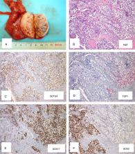
          The International Society of Urological Pathology (ISUP) organized a Consultation Conference in March 2019 dealing with applications of molecular pathology in Urogenital Pathology, including testicular tumors (with a focus on germ cell tumors [GCTs]), preceded by a survey among its members to get insight into current practices in testicular germ cell tumor (TGCT) diagnostics and adoption of the ISUP immunohistochemical guidelines published in 2014. On the basis of the premeeting survey, the most commonly used immunomarker panel includes OCT3/4, placental alkaline phosphate, D2-40, SALL4, CD117, and CD30 for GCTs and the documentation of germ cell neoplasia in situ (GCNIS). Molecular testing, specifically 12p copy gain, is informative to distinguish non-GCNIS versus GCNIS related GCTs, and establishing germ cell origin of tumors both in the context of primary and metastatic lesions. Other molecular methodologies currently available but not widely utilized for TGCTs include genome-wide and targeted approaches for specific genetic anomalies, P53 mutations, genomic MDM2 amplification, and detection of the p53 inactivating miR-371a-3p. The latter also holds promise as a serum marker for malignant TGCTs. This manuscript provides an update on the classification of TGCTs, and describes the current and future role of molecular-genetic testing. The following recommendations are made: (1) Presence of GCNIS should be documented in all cases along with extent of spermatogenesis; (2) Immunohistochemical staining is optional in the following scenarios: identification of GCNIS, distinguishing embryonal carcinoma from seminoma, confirming presence of yolk sac tumor and/or choriocarcinoma, and differentiating spermatocytic tumor from potential mimics; (3) Detection of gain of the short arm of chromosome 12 is diagnostic to differentiate between non-GCNIS versus GCNIS related GCTs and supportive to the germ cell origin of both primary and metastatic tumors.

          Related collections

          Most cited references98

          • Record: found
          • Abstract: found
          • Article: not found

          Testicular germ-cell tumours in a broader perspective.

          The germ-cell tumours are a fascinating group of neoplasms because of their unusual biology and the spectacular therapeutic results that have been obtained in these tumours. Traditionally, this group of neoplasms is presented in an organ-oriented approach. However, recent clinical and experimental data convincingly demonstrate that these neoplasms are one disease with separate entities that can manifest themselves in different anatomical sites. We propose five entities, in which the developmental potential is determined by the maturation stage and imprinting status of the originating germ cell. Recent progress begins to explain the apparent unpredictable development of germ-cell tumours and offers a basis for understanding their exquisite sensitivity to therapy.
            Bookmark
            • Record: found
            • Abstract: found
            • Article: not found

            POU5F1 (OCT3/4) identifies cells with pluripotent potential in human germ cell tumors.

            Human germ cell tumors (GCTs) may have variable histology and clinical behavior, depending on factors such as sex of the patient, age at clinical diagnosis, and anatomical site of the tumor. Some types of GCT, i.e., the seminomas/germinomas/dysgerminomas and embryonal carcinomas (the stem cell component of nonseminomas), have pluripotent potential, which is demonstrated by their capacity to differentiate into somatic and/or extraembryonic elements. Although embryonal carcinoma cells are intrinsically pluripotent, seminoma/germinoma/dysgerminoma cells, as well as their precursor carcinoma in situ/gonadoblastoma cells, have the phenotype of early germ cells that can be activated to pluripotency. The other types of GCT (teratomas and yolk sac tumors of infants and newborn, dermoid cyst of the ovary, and spermatocytic seminoma of elderly) are composed of (fully) differentiated tissues and lack the appearance of undifferentiated and pluripotent stem cells. OCT3/4, a transcription factor also known as OTF3 and POU5F1, is involved in regulation of pluripotency during normal development and is detectable in embryonic stem and germ cells. We analyzed the presence of POU5F1 in GCT and other tumor types using immunohistochemistry. The protein was consistently detected in carcinoma in situ/gonadoblastoma, seminomas/germinoma/dysgerminoma, and embryonal carcinoma but not in the various types of differentiated nonseminomas. Multitumor tissue microarray analysis covering >100 different tumor categories and 3600 individual cancers verified that POU5F1 expression is specific for particular subtypes of GCT of adults. No protein was observed in GCT of newborn and infants, spermatocytic seminomas, and the various tumors of nongerm cell origin. In addition, no difference in staining pattern was found in chemosensitive and chemoresistant GCT of adults. These results indicate preservation of the link between POU5F1 and pluripotency, as reported during normal development, after malignant transformation. Therefore, POU5F1 immunohistochemistry is an informative diagnostic tool for pluripotent GCT and offers new insights into the histological heterogeneity of this cancer.
              Bookmark
              • Record: found
              • Abstract: found
              • Article: found
              Is Open Access

              Serum Levels of MicroRNA-371a-3p (M371 Test) as a New Biomarker of Testicular Germ Cell Tumors: Results of a Prospective Multicentric Study

              PURPOSE Previous studies suggested that serum levels of microRNA (miR)-371a-3p (so-called M371 test) have a much higher sensitivity and specificity than the classic markers of testicular germ cell tumors (GCTs) and are applicable toward both seminoma and nonseminoma. We sought to confirm the usefulness of this test as a novel biomarker for GCT. PATIENTS AND METHODS In a prospective, multicentric study, serum samples of 616 patients with testicular GCTs and 258 male controls were examined for serum levels of miRNA-371a-3p (miR levels) by quantitative polymerase chain reaction. The GCT population encompassed 359 patients with seminoma and 257 with nonseminoma; 371 had clinical stage I disease, 201 had systemic disease, and 46 had relapses. Paired measurements before and after orchiectomy were performed in 424 patients; 118 with systemic disease had serial measurements during treatment. miR levels were compared with those of β-human chorionic gonadotropin, α-fetoprotein, and lactate dehydrogenase. RESULTS For the primary diagnosis of GCT, the M371 test showed a sensitivity of 90.1%, a specificity of 94.0%, an area under the curve of 0.966 upon receiver operating characteristic analysis, and a positive predictive value of 97.2%. α-Fetoprotein, β-human chorionic gonadotropin, and lactate dehydrogenase had sensitivities of less than 50% in seminoma and slightly higher sensitivities in nonseminomas. miR levels were significantly associated with clinical stage, primary tumor size, and response to treatment. Relapses had elevated miR levels that subsequently dropped to normal upon remission. Teratoma did not express miR-371a-3p. CONCLUSION The M371 test outperforms the classic markers of GCT with both a sensitivity and a specificity greater than 90%. All histologic subgroups, except teratoma, express this marker. The test could be considered for clinical implementation after further validation.
                Bookmark

                Author and article information

                Journal
                Am J Surg Pathol
                Am. J. Surg. Pathol
                PAS
                The American Journal of Surgical Pathology
                Wolters Kluwer Health, Inc
                0147-5185
                1532-0979
                July 2020
                20 March 2020
                : 44
                : 7
                : e66-e79
                Affiliations
                [* ]Princess Máxima Center for Pediatric Oncology, Utrecht, The Netherlands
                []Laboratory Medicine Program, University Health Network, Toronto, Canada
                Departments of []Pathology and Laboratory Medicine
                [# ]Pathology, Indiana University School of Medicine, Indianapolis, IN
                [§ ]Department of Oncology and Pathology, Karolinska Institutet Sweden, Solna, Sweden
                []Department of Pathology, University Hospital Bonn, Bonn, Germany
                []Department of Pathology, Stanford University School of Medicine, Stanford, CA
                Author notes
                Correspondence: Leendert H.J. Looijenga, PhD, Princess Máxima Center for Pediatric Oncology, Utrecht 3084 CS, The Netherlands (e-mail: l.looijenga@ 123456prinsesmaximacentrum.nl ).
                Article
                00004
                10.1097/PAS.0000000000001465
                7289140
                32205480
                d694e5d1-30ac-492b-afa0-3b418e2ea27c
                Copyright © 2020 The Author(s). Published by Wolters Kluwer Health, Inc.

                This is an open-access article distributed under the terms of the Creative Commons Attribution-Non Commercial-No Derivatives License 4.0 (CCBY-NC-ND), where it is permissible to download and share the work provided it is properly cited. The work cannot be changed in any way or used commercially without permission from the journal. http://creativecommons.org/licenses/by-nc-nd/4.0/

                History
                Categories
                Special Articles
                Custom metadata
                T
                TRUE
                ONLINE-ONLY

                testis,germ cell tumor,guidelines,immunohistochemistry,molecular test,genetic test

                Comments

                Comment on this article