29
views
0
recommends
+1 Recommend
0 collections
    0
    shares
      • Record: found
      • Abstract: found
      • Article: found
      Is Open Access

      Usefulness of Dermatoscopy for the Preoperative Assessment of the Histopathologic Aggressiveness of Basal Cell Carcinoma

      research-article

      Read this article at

      Bookmark
          There is no author summary for this article yet. Authors can add summaries to their articles on ScienceOpen to make them more accessible to a non-specialist audience.

          Abstract

          Background

          Limited information is available regarding dermatoscopic differences between non-aggressive and aggressive types of basal cell carcinoma (BCC).

          Objective

          To investigate dermatoscopic differences between non-aggressive and aggressive types.

          Methods

          We evaluated 145 histopathologically confirmed BCCs from 141 patients. Histopathologic types and aggressiveness from 4 mm punch biopsy and their dermatoscopic findings were evaluated. We assessed the statistical significance of dermatoscopic differences between non-aggressive and aggressive types. To objectively predict aggressiveness, we created a "dermatoscopic index of BCC aggressiveness" in which 1 point was added and subtracted for each dermatoscopic finding significantly higher in aggressive and non-aggressive types, respectively.

          Results

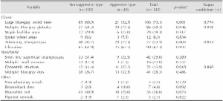
          Large blue-gray ovoid nests were found more frequently in non-aggressive type than aggressive one (85/105 [80.9%] vs. 21/40 [52.5%], p=0.001). Compared to non-aggressive type, aggressive type had more multiple blue-gray globules (29/40 [72.5%] vs. 57/105 [54.3%], p=0.046), arborizing telangiectasia (29/40 [72.5%] vs. 48/105 [45.7%], p=0.004), and concentric structure (11/40 [27.5%] vs. 12/105 [11.4%], p=0.018). Regarding dermatoscopic index, cases of aggressive type with a score of 1 were most common (n=18, 45.0%), followed by a score of 2 (n=14, 35.0%). Limited number of aggressive type of BCCs and the effect of width on the determination of histopathologic aggressiveness.

          Conclusion

          Aggressive type BCCs more often exhibited multiple blue-gray globules, arborizing telangiectasia, and concentric structure, while the non-aggressive type exhibited large blue-gray ovoid nests more frequently. Score exceeding 2 on the dermoscopic index can be screening criteria for aggressiveness. These dermatoscopic features and dermoscopic index could be useful for assessing aggressiveness of BCCs before surgery.

          Related collections

          Most cited references22

          • Record: found
          • Abstract: found
          • Article: not found

          Dermatoscopy of basal cell carcinoma: morphologic variability of global and local features and accuracy of diagnosis.

          Early detection of basal cell carcinoma (BCC) is crucial to reduce the morbidity of this tumor. We sought to investigate the variability and diagnostic significance of dermatoscopic features of BCCs. We conducted retrospective dermatoscopic analysis of 609 BCCs and 200 melanocytic and nonmelanocytic lesions, and assessment of interrater reliability of dermatoscopic BCC criteria. Lesions included nonpigmented (15.1%), lightly pigmented (33.2%), pigmented (42.7%), and heavily pigmented (9%) BCCs. Classic BCC patterns including arborizing telangiectasia (57.1%), blue/gray ovoid nests (47.5%), ulceration (39.2%), multiple blue/gray globules (26.1%), leaflike areas (15.9%), and spoke-wheel areas (9%) were significantly increased in pigmented BCCs compared with nonpigmented and heavily pigmented BCCs (P = .0001). Among nonclassic BCC patterns, we detected short fine superficial telangiectasia (10%) and multiple small erosions (8.5%), and described two new patterns named "concentric structures" (7.6%) and "multiple in-focus blue/gray dots" (5.1%). Dermatoscopic features suggestive of melanocytic lesions (eg, multiple brown to black dots/globules, blue/white veillike structures, and nonarborizing vessels) were observed in 40.6% BCCs and significantly increased in heavily pigmented BCCs (P < .0001). Expert observers provided an accurate (sensitivity: 97%) and reliable (K: 87%) dermatoscopic diagnosis of BCC, although a significant difference in terms of specificity (P = .0002) and positive predictive value (P = .0004) was found. Arborizing telangiectasia, leaflike areas, and large blue/gray ovoid nests represented reliable and robust diagnostic parameters. The study was retrospective. BCCs show a large spectrum of global and local dermatoscopic features; heavily pigmented BCCs show the most challenging combinations of dermatoscopic features.
            Bookmark
            • Record: found
            • Abstract: found
            • Article: not found

            Dermoscopy of pigmented skin lesions.

            Dermoscopy is an in vivo method for the early diagnosis of malignant melanoma and the differential diagnosis of pigmented lesions of the skin. It has been shown to increase diagnostic accuracy over clinical visual inspection in the hands of experienced physicians. This article is a review of the principles of dermoscopy as well as recent technological developments.
              Bookmark
              • Record: found
              • Abstract: found
              • Article: not found

              Surface microscopy of pigmented basal cell carcinoma.

              To describe the relevant morphologic features and to create a simple diagnostic method for pigmented basal cell carcinoma (BCC) using in vivo cutaneous surface microscopy (ie, dermoscopy, dermatoscopy, or oil epiluminescence microscopy). Pigmented skin lesions were photographed in vivo using immersion oil (surface microscopy). All pigmented skin lesions were excised and reviewed for histological diagnosis. Photographs of 142 pigmented BCCs, 142 invasive melanomas, and 142 benign pigmented skin lesions were randomly divided into 2 equally sized training and test sets. Images from the training set were scored for 45 surface microscopy features. From this a model was derived and tested on the independent test set. All patients were recruited from the primary case and referral centers of the Sydney Melanoma Unit, Sydney, Australia, and the Skin and Cancer Unit, Skin and Cancer Associates, Plantation, Fla. A random sample (selected from a larger database) of patients whose lesions were excised. Sensitivity and specificity of the model for diagnosis of pigmented BCCs. The following model was created. For a pigmented BCC to be diagnosed it must not have the negative feature of a pigment network and must have 1 or more of the following 6 positive features: large gray-blue ovoid nests, multiple gray-blue globules, maple leaflike areas, spoke wheel areas, ulceration, and arborizing "treelike" telangiectasia. On an independent test set the model had a sensitivity of 97% for the diagnosis of pigmented BCCs and a specificity of 93% for the invasive melanoma set and 92% for the benign pigmented skin lesion set. A robust surface microscopy method is described that allows the diagnosis of pigmented BCCs from invasive melanomas and benign pigmented skin lesions. Arch Dermatol. 2000;136:1012-1016
                Bookmark

                Author and article information

                Journal
                Ann Dermatol
                Ann Dermatol
                AD
                Annals of Dermatology
                Korean Dermatological Association; The Korean Society for Investigative Dermatology
                1013-9087
                2005-3894
                December 2015
                07 December 2015
                : 27
                : 6
                : 682-687
                Affiliations
                [1 ]Department of Dermatology, Pusan National University School of Medicine, Busan, Korea.
                [2 ]Biomedical Research Institute, Pusan National University Hospital, Busan, Korea.
                Author notes
                Corresponding author: Moon-Bum Kim, Department of Dermatology, Pusan National University School of Medicine, 305 Gudeok-ro, Seo-gu, Busan 49241, Korea. Tel: 82-51-240-7338, Fax: 82-51-245-9467, drkmp@ 123456hanmail.net
                Article
                10.5021/ad.2015.27.6.682
                4695419
                26719636
                ddb1238d-efb7-494f-b7e1-713dc5d760c7
                Copyright © 2015 The Korean Dermatological Association and The Korean Society for Investigative Dermatology

                This is an Open Access article distributed under the terms of the Creative Commons Attribution Non-Commercial License ( http://creativecommons.org/licenses/by-nc/4.0) which permits unrestricted non-commercial use, distribution, and reproduction in any medium, provided the original work is properly cited.

                History
                : 11 February 2014
                : 30 September 2014
                : 12 October 2014
                Categories
                Original Article

                Dermatology
                basal cell carcinoma,dermatoscopy,histopathologic aggressiveness
                Dermatology
                basal cell carcinoma, dermatoscopy, histopathologic aggressiveness

                Comments

                Comment on this article