7
views
0
recommends
+1 Recommend
0 collections
    0
    shares
      • Record: found
      • Abstract: found
      • Article: found
      Is Open Access

      Coronary Angioscopy Findings before and after Excimer Laser Coronary Angioplasty for Bare-Metal Stent In-Stent Restenosis

      case-report

      Read this article at

      Bookmark
          There is no author summary for this article yet. Authors can add summaries to their articles on ScienceOpen to make them more accessible to a non-specialist audience.

          Abstract

          A 59-year-old man who underwent bare-metal stent implantation in the left-anterior descending artery (LAD) 15 years previously was admitted to our hospital because of stable angina pectoris. Coronary angiography revealed 75% in-stent restenosis at the proximal LAD, showing a fractional flow reserve of 0.74 at the distal LAD (Figure 1). This lesion was successfully treated using excimer laser coronary angioplasty (ELCA) and Xience Alpine® (Abbott Vascular, Santa Clara, CA, USA) stent implantation under optical frequency domain imaging (OFDI) guidance (FastView®, Terumo, Tokyo, Japan) which demonstrated a cavity due to plaque rupture with thrombi, and fibroatheroma 
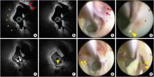
(Figure 2A and B). We performed coronary angioscopy (Forwardlooking®, OVALIS, Osaka, Japan) pre and post-ELCA for further evaluation with direct vision. Pre-ELCA coronary angioscopy showed a cavity due to plaque rupture with thrombi, indicating the progression of in-stent neoatherosclerosis (Figure 2C). Incomplete stent coverage was confirmed at the stent's proximal segment (Figure 2D). We performed ELCA 6 times using a 1.4-mm concentric laser catheter (CVX300®, Spectranetics, Colorado Springs, CO, USA) at a pulse rate 25 Hz, and energy output 45 mL/mm2. OFDI detected the ablation of in-stent surficial fibrous plaque after ELCA (Figure 2E and F). Coronary angioscopy revealed neointimal minor bleeding, and stent strut with neointima peeled off due to ELCA (Figure 2G and H). Final coronary angiography showed optimal results (Figure 3). Post-ELCA OFDI demonstrated that ablation of superficial plaque in in-stent area. Following OFDI, coronary angioscopy demonstrated surficial minor bleeding that was unclear on OFDI. Furthermore, coronary angioscopy clearly revealed the exposed strut after ELCA. Clinical studies using ELCA for in-stent restenosis have been reported1) 2); however, coronary angioscopy pre- and post-ELCA is unreported.

          Related collections

          Most cited references2

          • Record: found
          • Abstract: found
          • Article: not found

          Treatment of in-stent restenosis with excimer laser coronary angioplasty: benefits over scoring balloon angioplasty alone.

          Treatment of in-stent restenosis (ISR) is associated with a high incidence of recurrence. This study evaluated the clinical safety and 6-month efficacy of excimer laser coronary angioplasty (ELCA) before scoring balloon dilatation for the treatment of ISR. Twenty-three patients with ISR were included and treatment strategy of ISR was dependent on each operator. Twelve patients among those were treated with ELCA before scoring balloon dilatation (ELCA group) and 11 patients were treated with scoring balloon alone (non-ELCA group). Acute procedural results were evaluated by quantitative coronary angiography (QCA) and frequency domain optical coherence tomography (FD-OCT). Follow-up angiography was performed in all patients and the incidence of recurrent ISR and target lesion revascularization (TLR) was determined at 6 months after initial ISR treatment. Procedural success was achieved in all patients. Baseline clinical and angiographic characteristics were similar between groups. Maximum dilatation pressure of scoring balloon was significantly lower in the ELCA group than in the non-ELCA group (9.0 ± 3.1 vs. 14.9 ± 4.3 atm, p = 0.001). In follow-up angiography, the occurrence of TLR was similar between groups (16.7 vs. 45.5 %, p = 0.09), but the late luminal loss was significantly lower in the ELCA group (0.7 ± 0.6 vs. 1.3 ± 0.7 mm, p = 0.03). ELCA is a safe and feasible technique for the treatment of ISR and associated with a relatively low recurrent restenosis in comparison with scoring balloon dilatation alone.
            Bookmark
            • Record: found
            • Abstract: found
            • Article: not found

            The combined use of Drug-eluting balloon and Excimer laser for coronary artery Restenosis In-Stent Treatment: The DERIST study.

            Stents reduce angiographic restenosis in comparison with balloon angioplasty. The rate of in-stent restenosis (ISR), although less frequent than post-angioplasty restenosis, is becoming increasingly prevalent due to the recent exponential increase in the use of intracoronary stents. The aim of this study is to evaluate angiographic and clinical outcomes of PTCA in combination with the use of excimer laser coronary angioplasty (ELCA) and drug-eluting balloon (DEB) in treatment of in-stent restenosis (ISR).
              Bookmark

              Author and article information

              Journal
              Korean Circ J
              Korean Circ J
              KCJ
              Korean Circulation Journal
              The Korean Society of Cardiology
              1738-5520
              1738-5555
              May 2019
              04 April 2019
              : 49
              : 5
              : 465-467
              Affiliations
              [1 ]Department of Cardiology, Tokyo Metropolitan Hiroo Hospital, Tokyo, Japan.
              [2 ]Department of Cardiology, Japanese Red Cross Musashino Hospital, Tokyo, Japan.
              Author notes
              Correspondence to Shinichiro Masuda, MD. Department of Cardiology, Tokyo Metropolitan Hiroo Hospital, 2-34-10, Ebisu, Shibuya-ku, Tokyo 150-0013, Japan. giga627@ 123456yahoo.co.jp
              Author information
              https://orcid.org/0000-0002-2791-8538
              https://orcid.org/0000-0002-0676-5595
              https://orcid.org/0000-0002-3232-8014
              https://orcid.org/0000-0002-6923-9832
              Article
              10.4070/kcj.2019.0010
              6511532
              31074216
              df825cae-d248-4f46-96c6-b739796a93bd
              Copyright © 2019. The Korean Society of Cardiology

              This is an Open Access article distributed under the terms of the Creative Commons Attribution Non-Commercial License ( https://creativecommons.org/licenses/by-nc/4.0) which permits unrestricted noncommercial use, distribution, and reproduction in any medium, provided the original work is properly cited.

              History
              : 07 January 2019
              : 05 March 2019
              : 25 March 2019
              Categories
              Images in Cardiovascular Medicine

              Cardiovascular Medicine
              Cardiovascular Medicine

              Comments

              Comment on this article