4
views
0
recommends
+1 Recommend
0 collections
    0
    shares
      • Record: found
      • Abstract: found
      • Article: found
      Is Open Access

      High-Fat Diet-Induced Mitochondrial Dysfunction Promotes Genioglossus Injury – A Potential Mechanism for Obstructive Sleep Apnea with Obesity

      research-article

      Read this article at

      Bookmark
          There is no author summary for this article yet. Authors can add summaries to their articles on ScienceOpen to make them more accessible to a non-specialist audience.

          Abstract

          Purpose

          Obesity is a worldwide metabolic disease and a critical risk factor for several chronic conditions. Obstructive sleep apnea (OSA) is an important complication of obesity. With the soaring morbidity of obesity, the prevalence of OSA has markedly increased. However, the underlying mechanism of the high relevance between obesity and OSA has not been elucidated. This study investigated the effects of obesity on the structure and function of the genioglossus to explore the possible mechanisms involved in OSA combined with obesity.

          Methods

          Six-week-old male C57BL/6J mice were fed high-fat diet (HFD, 60% energy) or normal diet (Control, 10% energy) for 16 weeks. The muscle fibre structure and electromyography (EMG) activity of genioglossus were measured. The ultrastructure and function of mitochondrial, oxidative damage and apoptosis in genioglossus were detected by transmission electron microscopy (TEM), qPCR, Western blotting, immunohistochemistry and TUNEL staining. We further studied the influence of palmitic acid (PA) on the proliferation and myogenic differentiation of C2C12 myoblasts, as well as mitochondrial function, oxidative stress, and apoptosis in C2C12 myotubes.

          Results

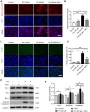
          Compared with the control, the number of muscle fibres was decreased, the fibre type was remarkably changed, and the EMG activity had declined in genioglossus. In addition, a HFD also reduced mitochondria quantity and function, induced excessive oxidative stress and increased apoptosis in genioglossus. In vitro, PA treatment significantly inhibited the proliferation and myogenic differentiation of C2C12 myoblasts. Moreover, PA decreased the mitochondrial membrane potential, upregulated mitochondrial reactive oxygen species (ROS) levels, and activated the mitochondrial-related apoptotic pathway in myotubes.

          Conclusion

          Our findings suggest that a HFD caused genioglossus injury in obese mice. The mitochondrial dysfunction and the accompanying oxidative stress were involved in the genioglossus injury, which may provide potential therapeutic targets for OSA with obesity.

          Related collections

          Most cited references49

          • Record: found
          • Abstract: found
          • Article: not found

          Mitochondria as multifaceted regulators of cell death

          Through their many and varied metabolic functions, mitochondria power life. Paradoxically, mitochondria also have a central role in apoptotic cell death. Upon induction of mitochondrial apoptosis, mitochondrial outer membrane permeabilization (MOMP) usually commits a cell to die. Apoptotic signalling downstream of MOMP involves cytochrome c release from mitochondria and subsequent caspase activation. As such, targeting MOMP in order to manipulate cell death holds tremendous therapeutic potential across different diseases, including neurodegenerative diseases, autoimmune disorders and cancer. In this Review, we discuss new insights into how mitochondria regulate apoptotic cell death. Surprisingly, recent data demonstrate that besides eliciting caspase activation, MOMP engages various pro-inflammatory signalling functions. As we highlight, together with new findings demonstrating cell survival following MOMP, this pro-inflammatory role suggests that mitochondria-derived signalling downstream of pro-apoptotic cues may also have non-lethal functions. Finally, we discuss the importance and roles of mitochondria in other forms of regulated cell death, including necroptosis, ferroptosis and pyroptosis. Collectively, these new findings offer exciting, unexplored opportunities to target mitochondrial regulation of cell death for clinical benefit.
            Bookmark
            • Record: found
            • Abstract: found
            • Article: not found

            Increased prevalence of sleep-disordered breathing in adults.

            Sleep-disordered breathing is a common disorder with a range of harmful sequelae. Obesity is a strong causal factor for sleep-disordered breathing, and because of the ongoing obesity epidemic, previous estimates of sleep-disordered breathing prevalence require updating. We estimated the prevalence of sleep-disordered breathing in the United States for the periods of 1988-1994 and 2007-2010 using data from the Wisconsin Sleep Cohort Study, an ongoing community-based study that was established in 1988 with participants randomly selected from an employed population of Wisconsin adults. A total of 1,520 participants who were 30-70 years of age had baseline polysomnography studies to assess the presence of sleep-disordered breathing. Participants were invited for repeat studies at 4-year intervals. The prevalence of sleep-disordered breathing was modeled as a function of age, sex, and body mass index, and estimates were extrapolated to US body mass index distributions estimated using data from the National Health and Nutrition Examination Survey. The current prevalence estimates of moderate to severe sleep-disordered breathing (apnea-hypopnea index, measured as events/hour, ≥15) are 10% (95% confidence interval (CI): 7, 12) among 30-49-year-old men; 17% (95% CI: 15, 21) among 50-70-year-old men; 3% (95% CI: 2, 4) among 30-49-year-old women; and 9% (95% CI: 7, 11) among 50-70 year-old women. These estimated prevalence rates represent substantial increases over the last 2 decades (relative increases of between 14% and 55% depending on the subgroup).
              Bookmark
              • Record: found
              • Abstract: found
              • Article: not found

              Fiber types in mammalian skeletal muscles.

              Mammalian skeletal muscle comprises different fiber types, whose identity is first established during embryonic development by intrinsic myogenic control mechanisms and is later modulated by neural and hormonal factors. The relative proportion of the different fiber types varies strikingly between species, and in humans shows significant variability between individuals. Myosin heavy chain isoforms, whose complete inventory and expression pattern are now available, provide a useful marker for fiber types, both for the four major forms present in trunk and limb muscles and the minor forms present in head and neck muscles. However, muscle fiber diversity involves all functional muscle cell compartments, including membrane excitation, excitation-contraction coupling, contractile machinery, cytoskeleton scaffold, and energy supply systems. Variations within each compartment are limited by the need of matching fiber type properties between different compartments. Nerve activity is a major control mechanism of the fiber type profile, and multiple signaling pathways are implicated in activity-dependent changes of muscle fibers. The characterization of these pathways is raising increasing interest in clinical medicine, given the potentially beneficial effects of muscle fiber type switching in the prevention and treatment of metabolic diseases.
                Bookmark

                Author and article information

                Journal
                Nat Sci Sleep
                Nat Sci Sleep
                nss
                Nature and Science of Sleep
                Dove
                1179-1608
                23 December 2021
                2021
                : 13
                : 2203-2219
                Affiliations
                [1 ]Department of Orthodontics, Shanghai Stomatological Hospital, Fudan University , Shanghai, People’s Republic of China
                [2 ]Shanghai Key Laboratory of Craniomaxillofacial Development and Diseases, Fudan University , Shanghai, People’s Republic of China
                [3 ]Department of Periodontology, Shanghai Stomatological Hospital, Fudan University , Shanghai, People’s Republic of China
                Author notes
                Correspondence: Yuehua Liu; Liming Yu Shanghai Stomatological Hospital, Fudan University , No. 356 East Beijing Road, Shanghai, 200001, People’s Republic of China Tel +86 2163298475 Fax +86 2163614515 Email liuyuehua@fudan.edu.cn; ylmphoebe@126.com
                Author information
                http://orcid.org/0000-0001-5385-7452
                http://orcid.org/0000-0001-8029-0711
                Article
                343721
                10.2147/NSS.S343721
                8711738
                34992480
                dfcb32ea-7f4c-4442-a307-8a9dd6e63eb9
                © 2021 Chen et al.

                This work is published and licensed by Dove Medical Press Limited. The full terms of this license are available at https://www.dovepress.com/terms.php and incorporate the Creative Commons Attribution – Non Commercial (unported, v3.0) License ( http://creativecommons.org/licenses/by-nc/3.0/). By accessing the work you hereby accept the Terms. Non-commercial uses of the work are permitted without any further permission from Dove Medical Press Limited, provided the work is properly attributed. For permission for commercial use of this work, please see paragraphs 4.2 and 5 of our Terms ( https://www.dovepress.com/terms.php).

                History
                : 10 October 2021
                : 14 December 2021
                Page count
                Figures: 8, References: 49, Pages: 17
                Funding
                Funded by: the National Science Foundation of Shanghai;
                Funded by: the Youth Program of Shanghai Municipal Health and Family Planning Commission grant;
                This project was supported by NSFC grants 81771109 and 82071153 to Yuehua Liu, 81901031 to Xinxin Han and 81701011 to Bingjiao Zhao; the National Science Foundation of Shanghai (grant no. 19ZR1445400 to Xinxin Han); the Youth Program of Shanghai Municipal Health and Family Planning Commission grant (grant no. 20184Y0228 to Qingqing Chen).
                Categories
                Original Research

                obesity,obstructive sleep apnea,genioglossus,muscle,mitochondria

                Comments

                Comment on this article