3
views
0
recommends
+1 Recommend
0 collections
    0
    shares
      • Record: found
      • Abstract: found
      • Article: found
      Is Open Access

      Combination of neuron-specific enolase measurement and initial neurological examination for the prediction of neurological outcomes after cardiac arrest

      research-article

      Read this article at

      Bookmark
          There is no author summary for this article yet. Authors can add summaries to their articles on ScienceOpen to make them more accessible to a non-specialist audience.

          Abstract

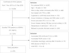
          This study aimed to investigate the efficacy of the combination of neuron-specific enolase (NSE) measurement and initial neurological examination in predicting the neurological outcomes of patients with cardiac arrest (CA) by retrospectively analyzing data from the Korean Hypothermia Network prospective registry. NSE levels were recorded at 48 and 72 h after CA. The initial Full Outline of UnResponsiveness (FOUR) and Glasgow Coma Scale (GCS) scores were recorded. These variables were categorized using the scorecard method. The primary endpoint was poor neurological outcomes at 6 months. Of the 475 patients, 171 (36%) had good neurological outcomes at 6 months. The areas under the curve (AUCs) of the categorized NSE levels at 72 h, GCS score, and FOUR score were 0.889, 0.722, and 0.779, respectively. The AUCs of the combinations of categorized NSE levels at 72 h with categorized GCS scores and FOUR score were 0.910 and 0.912, respectively. Each combination was significantly higher than the AUC value of the categorized NSE level at 72 h alone (with GCS: p = 0.015; with FOUR: p = 0.026). Combining NSE measurement and initial neurological examination improved the prediction of neurological outcomes.

          Related collections

          Most cited references30

          • Record: found
          • Abstract: not found
          • Article: not found

          Part 8: Post-Cardiac Arrest Care: 2015 American Heart Association Guidelines Update for Cardiopulmonary Resuscitation and Emergency Cardiovascular Care.

            Bookmark
            • Record: found
            • Abstract: found
            • Article: not found

            Part 9: post-cardiac arrest care: 2010 American Heart Association Guidelines for Cardiopulmonary Resuscitation and Emergency Cardiovascular Care.

            The goal of immediate post-cardiac arrest care is to optimize systemic perfusion, restore metabolic homeostasis, and support organ system function to increase the likelihood of intact neurological survival. The post-cardiac arrest period is often marked by hemodynamic instability as well as metabolic abnormalities. Support and treatment of acute myocardial dysfunction and acute myocardial ischemia can increase the probability of survival. Interventions to reduce secondary brain injury, such as therapeutic hypothermia, can improve survival and neurological recovery. Every organ system is at risk during this period, and patients are at risk of developing multiorgan dysfunction. The comprehensive treatment of diverse problems after cardiac arrest involves multidisciplinary aspects of critical care, cardiology, and neurology. For this reason, it is important to admit patients to appropriate critical-care units with a prospective plan of care to anticipate, monitor, and treat each of these diverse problems. It is also important to appreciate the relative strengths and weaknesses of different tools for estimating the prognosis of patients after cardiac arrest.
              Bookmark
              • Record: found
              • Abstract: found
              • Article: found
              Is Open Access

              Prognostication after cardiac arrest

              Hypoxic–ischaemic brain injury (HIBI) is the main cause of death in patients who are comatose after resuscitation from cardiac arrest. A poor neurological outcome—defined as death from neurological cause, persistent vegetative state, or severe neurological disability—can be predicted in these patients by assessing the severity of HIBI. The most commonly used indicators of severe HIBI include bilateral absence of corneal and pupillary reflexes, bilateral absence of N2O waves of short-latency somatosensory evoked potentials, high blood concentrations of neuron specific enolase, unfavourable patterns on electroencephalogram, and signs of diffuse HIBI on computed tomography or magnetic resonance imaging of the brain. Current guidelines recommend performing prognostication no earlier than 72 h after return of spontaneous circulation in all comatose patients with an absent or extensor motor response to pain, after having excluded confounders such as residual sedation that may interfere with clinical examination. A multimodal approach combining multiple prognostication tests is recommended so that the risk of a falsely pessimistic prediction is minimised.
                Bookmark

                Author and article information

                Contributors
                suka1212@hanmail.net
                Journal
                Sci Rep
                Sci Rep
                Scientific Reports
                Nature Publishing Group UK (London )
                2045-2322
                23 July 2021
                23 July 2021
                2021
                : 11
                : 15067
                Affiliations
                [1 ]GRID grid.255166.3, ISNI 0000 0001 2218 7142, Department of Emergency Medicine, , Dong-A University College of Medicine, ; Busan, South Korea
                [2 ]GRID grid.264381.a, ISNI 0000 0001 2181 989X, Department of Emergency Medicine, Samsung Changwon Hospital, , Sungkyunkwan University School of Medicine, ; 158, Paryong-ro, Masanhoewon-gu, Changwon-si, Gyeongsangnam-do 630-723 South Korea
                [3 ]GRID grid.411947.e, ISNI 0000 0004 0470 4224, Department of Emergency Medicine, Seoul St. Mary’s Hospital, College of Medicine, , The Catholic University of Korea, ; Seoul, South Korea
                [4 ]GRID grid.411947.e, ISNI 0000 0004 0470 4224, Department of Emergency Medicine, Bucheon St. Mary’s Hospital, College of Medicine, , The Catholic University of Korea, ; Seoul, South Korea
                [5 ]GRID grid.264381.a, ISNI 0000 0001 2181 989X, Department of Emergency Medicine, Samsung Medical Center, , Sungkyunkwan University School of Medicine, ; Seoul, South Korea
                [6 ]GRID grid.14005.30, ISNI 0000 0001 0356 9399, Department of Emergency Medicine, , Chonnam National University, Chonnam National University Hospital, ; Gwangju, South Korea
                Article
                94555
                10.1038/s41598-021-94555-0
                8302684
                34302037
                e0f2139c-3f16-4793-9e02-42b06f670baa
                © The Author(s) 2021

                Open Access This article is licensed under a Creative Commons Attribution 4.0 International License, which permits use, sharing, adaptation, distribution and reproduction in any medium or format, as long as you give appropriate credit to the original author(s) and the source, provide a link to the Creative Commons licence, and indicate if changes were made. The images or other third party material in this article are included in the article's Creative Commons licence, unless indicated otherwise in a credit line to the material. If material is not included in the article's Creative Commons licence and your intended use is not permitted by statutory regulation or exceeds the permitted use, you will need to obtain permission directly from the copyright holder. To view a copy of this licence, visit http://creativecommons.org/licenses/by/4.0/.

                History
                : 15 January 2021
                : 13 July 2021
                Categories
                Article
                Custom metadata
                © The Author(s) 2021

                Uncategorized
                medical research,neurology
                Uncategorized
                medical research, neurology

                Comments

                Comment on this article