3
views
0
recommends
+1 Recommend
0 collections
    0
    shares
      • Record: found
      • Abstract: found
      • Article: found
      Is Open Access

      A CT-based radiomics approach to predict intra-tumoral tertiary lymphoid structures and recurrence of intrahepatic cholangiocarcinoma

      research-article

      Read this article at

      Bookmark
          There is no author summary for this article yet. Authors can add summaries to their articles on ScienceOpen to make them more accessible to a non-specialist audience.

          Abstract

          Purpose

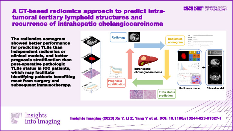
          To predict the tertiary lymphoid structures (TLSs) status and recurrence-free survival (RFS) of intrahepatic cholangiocarcinoma (ICC) patients using preoperative CT radiomics.

          Patients and methods

          A total of 116 ICC patients were included (training: 86; external validation: 30). The enhanced CT images were performed for the radiomics model. The logistic regression analysis was applied for the clinical model. The combined model was based on the clinical and radiomics models.

          Results

          A total of 107 radiomics features were extracted, and after being eliminated and selected, six features were combined to establish a radiomics model for TLSs prediction. Arterial phase diffuse hyperenhancement and AJCC 8th stage were combined to construct a clinical model. The combined (radiomics nomogram) model outperformed both the independent radiomics model and clinical model in the training cohort (AUC, 0.85 vs. 0.82 and 0.75, respectively) and was validated in the external validation cohort (AUC, 0.88 vs. 0.86 and 0.71, respectively). Patients in the rad-score no less than −0.76 (low-risk) group showed significantly better RFS than those in the less than −0.76 (high-risk) group ( p < 0.001, C-index = 0.678). Patients in the nomogram score no less than −1.16 (low-risk) group showed significantly better RFS than those of the less than −1.16 (high-risk) group ( p < 0.001, C-index = 0.723).

          Conclusions

          CT radiomics nomogram could serve as a preoperative biomarker of intra-tumoral TLSs status, better than independent radiomics or clinical models; preoperative CT radiomics nomogram achieved accurate stratification for RFS of ICC patients, better than the postoperative pathologic TLSs status.

          Critical relevance statement

          The radiomics nomogram showed better performance in predicting TLSs than independent radiomics or clinical models and better prognosis stratification than postoperative pathologic TLSs status in ICC patients, which may facilitate identifying patients benefiting most from surgery and subsequent immunotherapy.

          Key points

          • The combined (radiomics nomogram) model consisted of the radiomics model and clinical model (arterial phase diffuse hyperenhancement and AJCC 8th stage).

          • The radiomics nomogram showed better performance in predicting TLSs than independent radiomics or clinical models in ICC patients.

          • Preoperative CT radiomics nomogram achieved more accurate stratification for RFS of ICC patients than the postoperative pathologic TLSs status.

          Graphical Abstract

          Supplementary Information

          The online version contains supplementary material available at 10.1186/s13244-023-01527-1.

          Related collections

          Most cited references44

          • Record: found
          • Abstract: found
          • Article: not found

          The meaning and use of the area under a receiver operating characteristic (ROC) curve.

          A representation and interpretation of the area under a receiver operating characteristic (ROC) curve obtained by the "rating" method, or by mathematical predictions based on patient characteristics, is presented. It is shown that in such a setting the area represents the probability that a randomly chosen diseased subject is (correctly) rated or ranked with greater suspicion than a randomly chosen non-diseased subject. Moreover, this probability of a correct ranking is the same quantity that is estimated by the already well-studied nonparametric Wilcoxon statistic. These two relationships are exploited to (a) provide rapid closed-form expressions for the approximate magnitude of the sampling variability, i.e., standard error that one uses to accompany the area under a smoothed ROC curve, (b) guide in determining the size of the sample required to provide a sufficiently reliable estimate of this area, and (c) determine how large sample sizes should be to ensure that one can statistically detect differences in the accuracy of diagnostic techniques.
            Bookmark
            • Record: found
            • Abstract: found
            • Article: not found

            Radiomics: Images Are More than Pictures, They Are Data

            This report describes the process of radiomics, its challenges, and its potential power to facilitate better clinical decision making, particularly in the care of patients with cancer.
              Bookmark
              • Record: found
              • Abstract: found
              • Article: not found

              Radiomics: the bridge between medical imaging and personalized medicine

              Radiomics, the high-throughput mining of quantitative image features from standard-of-care medical imaging that enables data to be extracted and applied within clinical-decision support systems to improve diagnostic, prognostic, and predictive accuracy, is gaining importance in cancer research. Radiomic analysis exploits sophisticated image analysis tools and the rapid development and validation of medical imaging data that uses image-based signatures for precision diagnosis and treatment, providing a powerful tool in modern medicine. Herein, we describe the process of radiomics, its pitfalls, challenges, opportunities, and its capacity to improve clinical decision making, emphasizing the utility for patients with cancer. Currently, the field of radiomics lacks standardized evaluation of both the scientific integrity and the clinical relevance of the numerous published radiomics investigations resulting from the rapid growth of this area. Rigorous evaluation criteria and reporting guidelines need to be established in order for radiomics to mature as a discipline. Herein, we provide guidance for investigations to meet this urgent need in the field of radiomics.
                Bookmark

                Author and article information

                Contributors
                dr_fengye_ncc@163.com
                zhoujx888@126.com
                jmying@cicams.ac.cn
                zhaohong@cicams.ac.cn
                zhaoxinming@cicams.ac.cn
                Journal
                Insights Imaging
                Insights Imaging
                Insights into Imaging
                Springer Vienna (Vienna )
                1869-4101
                15 October 2023
                15 October 2023
                December 2023
                : 14
                : 173
                Affiliations
                [1 ]Department of Diagnostic Radiology, National Cancer Center/National Clinical Research Center for Cancer/Cancer Hospital, Chinese Academy of Medical Sciences and Peking Union Medical College, ( https://ror.org/02drdmm93) Beijing, China
                [2 ]Department of Pathology, National Cancer Center/National Clinical Research Center for Cancer/Cancer Hospital, Chinese Academy of Medical Sciences and Peking Union Medical College, ( https://ror.org/02drdmm93) Beijing, China
                [3 ] Department of Hepatobiliary Surgery, National Cancer Center/National Clinical Research Center for Cancer/Cancer Hospital, Chinese Academy of Medical Sciences and Peking Union Medical College, ( https://ror.org/02drdmm93) Beijing, China
                [4 ]Key Laboratory of Gene Editing Screening and Research and Development (R&D) of Digestive System Tumor Drugs, Chinese Academy of Medical Sciences and Peking Union Medical College, ( https://ror.org/02drdmm93) Beijing, China
                [5 ]GRID grid.414008.9, ISNI 0000 0004 1799 4638, Department of Hepatobiliary and Pancreatic Surgery, , Affiliated Cancer Hospital of Zhengzhou University, ; Zhengzhou, Henan China
                [6 ]Magnetic Resonance Imaging Research, General Electric Healthcare, Beijing, China
                Article
                1527
                10.1186/s13244-023-01527-1
                10577112
                37840098
                e1cecf9e-8476-4294-9375-f01a3cb6b78a
                © European Society of Radiology (ESR) 2023

                Open Access This article is licensed under a Creative Commons Attribution 4.0 International License, which permits use, sharing, adaptation, distribution and reproduction in any medium or format, as long as you give appropriate credit to the original author(s) and the source, provide a link to the Creative Commons licence, and indicate if changes were made. The images or other third party material in this article are included in the article's Creative Commons licence, unless indicated otherwise in a credit line to the material. If material is not included in the article's Creative Commons licence and your intended use is not permitted by statutory regulation or exceeds the permitted use, you will need to obtain permission directly from the copyright holder. To view a copy of this licence, visit http://creativecommons.org/licenses/by/4.0/.

                History
                : 4 July 2023
                : 13 September 2023
                Funding
                Funded by: FundRef http://dx.doi.org/10.13039/501100012166, National Key Research and Development Program of China;
                Award ID: 2020AAA0109503
                Award Recipient :
                Funded by: FundRef http://dx.doi.org/10.13039/501100001809, National Natural Science Foundation of China;
                Award ID: 81972311
                Award ID: 82141127
                Award Recipient :
                Funded by: CAMS Innovation Fund for Medical Sciences (CIFMS)
                Award ID: 2022-I2M-C&T-B-081
                Award Recipient :
                Funded by: Special Research Fund for Central Universities, Peking Union Medical College
                Award ID: 3332022026
                Award Recipient :
                Categories
                Original Article
                Custom metadata
                © European Society of Radiology (ESR) 2023

                Radiology & Imaging
                tertiary lymphoid structures,intrahepatic cholangiocarcinoma,radiomics,ct,recurrence

                Comments

                Comment on this article