34
views
0
recommends
+1 Recommend
0 collections
    0
    shares
      • Record: found
      • Abstract: found
      • Article: found
      Is Open Access

      Five-fraction CyberKnife radiotherapy for large brain metastases in critical areas: impact on the surrounding brain volumes circumscribed with a single dose equivalent of 14 Gy (V14) to avoid radiation necrosis

      research-article

      Read this article at

      Bookmark
          There is no author summary for this article yet. Authors can add summaries to their articles on ScienceOpen to make them more accessible to a non-specialist audience.

          Abstract

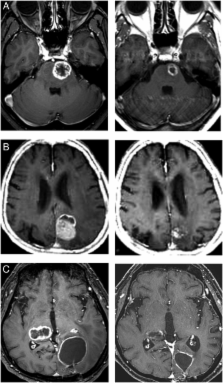
          The efficacy and toxicity of five-fraction CyberKnife radiotherapy were evaluated in patients with large brain metastases in critical areas. A total of 85 metastases in 78 patients, including tumors >30 cm 3 (4 cm in diameter) were treated with five-fraction CyberKnife radiotherapy with a median marginal dose of 31 Gy at a median prescribed isodose of 58%. Changes in the neurological manifestations, local tumor control, and adverse effects were investigated after treatment. The surrounding brain volumes circumscribed with 28.8 Gy (single dose equivalent to 14 Gy: V14) were measured to evaluate the risk of radiation necrosis. Neurological manifestations, such as motor weakness, visual disturbances and aphasia improved in 28 of 55 patients (50.9%). Local tumor control was obtained in 79 of 85 metastases (92.9%) during a median follow-up of eight months. Symptomatic edema occurred in 10 patients, and two of them (2.6%) required surgical resection because of radiation necrosis. The V14 of these patients was 3.0–19.7 cm 3. There were 16 lesions with a V14 of ≥7.0 cm 3, and two of these lesions developed extensive brain edema due to radiation necrosis. None of the patients with a V14 of <7.0 cm 3 exhibited edema requiring surgical intervention. We therefore conclude that a high rate of local tumor control and low rates of complications can be obtained after five-fraction CyberKnife radiotherapy for large metastases in critical areas. The V14 of the surrounding brain is therefore a useful indicator for the risk of radiation necrosis in patients with large metastases.

          Related collections

          Most cited references32

          • Record: found
          • Abstract: found
          • Article: not found

          Recursive partitioning analysis (RPA) of prognostic factors in three Radiation Therapy Oncology Group (RTOG) brain metastases trials.

          Promising results from new approaches such as radiosurgery or stereotactic surgery of brain metastases have recently been reported. Are these results due to the therapy alone or can the results be attributed in part to patient selection? An analysis of tumor/patient characteristics and treatment variables in previous Radiation Therapy Oncology Group (RTOG) brain metastases studies was considered necessary to fully evaluate the benefit of these new interventions. The database included 1200 patients from three consecutive RTOG trials conducted between 1979 and 1993, which tested several different dose fractionation schemes and radiation sensitizers. Using recursive partitioning analysis (RPA), a statistical methodology which creates a regression tree according to prognostic significance, eighteen pretreatment characteristics and three treatment-related variables were analyzed. According to the RPA tree the best survival (median: 7.1 months) was observed in patients or = 70, < 65 years of age with controlled primary and no extracranial metastases; Class 3: KPS < 70; Class 2- all others. Using these classes or stages, new treatment techniques can be tested on homogeneous patient groups.
            Bookmark
            • Record: found
            • Abstract: not found
            • Article: not found

            An overview of hypofractionation and introduction to this issue of seminars in radiation oncology.

              Bookmark
              • Record: found
              • Abstract: found
              • Article: not found

              Three-staged stereotactic radiotherapy without whole brain irradiation for large metastatic brain tumors.

              To evaluate the efficacy and toxicity of staged stereotactic radiotherapy with a 2-week interfraction interval for unresectable brain metastases more than 10 cm(3) in volume. Subjects included 43 patients (24 men and 19 women), ranging in age from 41 to 84 years, who had large brain metastases (> 10 cc in volume). Primary tumors were in the colon in 14 patients, lung in 12, breast in 11, and other in 6. The peripheral dose was 10 Gy in three fractions. The interval between fractions was 2 weeks. The mean tumor volume before treatment was 17.6 +/- 6.3 cm(3) (mean +/- SD). Mean follow-up interval was 7.8 months. The local tumor control rate, as well as overall, neurological, and qualitative survivals, were calculated using the Kaplan-Meier method. At the time of the second and third fractions, mean tumor volumes were 14.3 +/- 6.5 (18.8% reduction) and 10.6 +/- 6.1 cm(3) (39.8% reduction), respectively, showing significant reductions. The median overall survival period was 8.8 months. Neurological and qualitative survivals at 12 months were 81.8% and 76.2%, respectively. Local tumor control rates were 89.8% and 75.9% at 6 and 12 months, respectively. Tumor recurrence-free and symptomatic edema-free rates at 12 months were 80.7% and 84.4%, respectively. The 2-week interval allowed significant reduction of the treatment volume. Our results suggest staged stereotactic radiotherapy using our protocol to be a possible alternative for treating large brain metastases.
                Bookmark

                Author and article information

                Journal
                J Radiat Res
                J. Radiat. Res
                jrr
                jrr
                Journal of Radiation Research
                Oxford University Press
                0449-3060
                1349-9157
                March 2014
                1 November 2013
                1 November 2013
                : 55
                : 2
                : 334-342
                Affiliations
                [1 ]Cyber Center, Kanto Neurosurgical Hospital, 1120 Dai, Kumagaya, Saitama, 360-0804, Japan
                [2 ]Gunma University Heavy-ion Medical Research Center, 3-39-22 Showa-machi, Maebashi, Gunma, 371-8511, Japan
                [3 ]Department of Radiation Oncology, Gunma University Graduate School of Medicine, 3-39-22 Showa-machi, Maebashi, Gunma, 371-8511, Japan
                Author notes
                [* ]Corresponding author. Cyber Center, Kanto Neurosurgical Hospital, 1120 Dai, Kumagaya, Saitama, 360-0804, Japan. Tel: +81-485-21-3133; Fax: +81-485-24-6190; Email: chair.ino@ 123456instno-med.org .
                Article
                rrt127
                10.1093/jrr/rrt127
                3951086
                24187332
                e211f382-7959-486f-829e-c8311b50de9a
                © The Author 2013. Published by Oxford University Press on behalf of The Japan Radiation Research Society and Japanese Society for Therapeutic Radiology and Oncology.

                This is an Open Access article distributed under the terms of the Creative Commons Attribution License ( http://creativecommons.org/licenses/by/3.0/), which permits unrestricted reuse, distribution, and reproduction in any medium, provided the original work is properly cited.

                History
                : 24 June 2013
                : 23 August 2013
                : 18 September 2013
                Page count
                Pages: 21
                Categories
                Oncology

                Oncology & Radiotherapy
                large brain metastases,hypofractionated radiotherapy,five-session radiosurgery,radiation necrosis,v14

                Comments

                Comment on this article