1
views
0
recommends
+1 Recommend
0 collections
    0
    shares
      • Record: found
      • Abstract: found
      • Article: found
      Is Open Access

      Evaluation of exposure dose in fetal computed tomography using organ-effective modulation

      research-article

      Read this article at

      Bookmark
          There is no author summary for this article yet. Authors can add summaries to their articles on ScienceOpen to make them more accessible to a non-specialist audience.

          Abstract

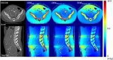
          Organ-effective modulation (OEM) is a computed tomography scanning technique that reduces the exposure dose to organs at risk. Ultrasonography is commonly used for prenatal imaging, but its reliability is reported to be limited. Radiography and computed tomography (CT) are reliable but pose risk of radiation exposure to the pregnant woman and her fetus. Although there are many reports on the exposure dose associated with fetal CT scans, no reports exist on OEM use in fetal CT scans. We measured the basic characteristics of organ-effective modulation (X-ray output modulation angle, maximum X-ray output modulation rate, total X-ray output modulation rate, and noise modulation) and used them in a Monte Carlo simulation to evaluate the effect of this technique on fetal CT scans in terms of image quality and exposure dose to the pregnant woman and fetus. Using ImPACT MC software, Monte Carlo simulations of OEM ON and OEM OFF were run on 8 cases involving fetal CT scans. We confirmed that the organ-effective modulation X-ray output modulation angle was 160°; the X-ray output modulation rate increased with increasing tube current; and no modulation occurred at tube currents of 80 mA or below. Our findings suggest that OEM has only a minimal effect in reducing organ exposure in pregnant women; therefore, it should be used on the anterior side (OEM ON,front) to reduce the exposure dose to the fetus.

          Related collections

          Most cited references26

          • Record: found
          • Abstract: not found
          • Article: not found

          Semiempirical model for generating tungsten target x-ray spectra

            Bookmark
            • Record: found
            • Abstract: found
            • Article: not found

            Validation of a Monte Carlo tool for patient-specific dose simulations in multi-slice computed tomography.

            Estimating the dose delivered to the patient in X-ray computed tomography (CT) examinations is not a trivial task. Monte Carlo (MC) methods appear to be the method of choice to assess the 3D dose distribution. The purpose of this work was to extend an existing MC-based tool to account for arbitrary scanners and scan protocols such as multi-slice CT (MSCT) scanners and to validate the tool in homogeneous and heterogeneous phantoms. The tool was validated by measurements on MSCT scanners for different scan protocols under known conditions. Quantitative CT Dose Index (CTDI) measurements were performed in cylindrical CTDI phantoms and in anthropomorphic thorax phantoms of various sizes; dose profiles were measured with thermoluminescent dosimeters (TLD) in the CTDI phantoms and compared with the computed dose profiles. The in-plane dose distributions were simulated and compared with TLD measurements in an Alderson-Rando phantom. The calculated dose values were generally within 10% of measurements for all phantoms and all investigated conditions. Three-dimensional dose distributions can be accurately calculated with the MC tool for arbitrary scanners and protocols including tube current modulation schemes. The use of the tool has meanwhile also been extended to further scanners and to flat-detector CT.
              Bookmark
              • Record: found
              • Abstract: found
              • Article: not found

              Radiation dose to the fetus for pregnant patients undergoing multidetector CT imaging: Monte Carlo simulations estimating fetal dose for a range of gestational age and patient size.

              To use Monte Carlo simulations of a current-technology multidetector computed tomographic (CT) scanner to investigate fetal radiation dose resulting from an abdominal and pelvic examination for a range of actual patient anatomies that include variation in gestational age and maternal size. Institutional review board approval was obtained for this HIPAA-compliant retrospective study. Twenty-four models of maternal and fetal anatomy were created from image data from pregnant patients who had previously undergone clinically indicated CT examination. Gestational age ranged from less than 5 weeks to 36 weeks. Simulated helical scans of the abdominal and pelvic region were performed, and a normalized dose (in milligrays per 100 mAs) was calculated for each fetus. Stepwise multiple linear regression was performed to analyze the correlation of dose with gestational age and anatomic measurements of maternal size and fetal location. Results were compared with several existing fetal dose estimation methods. Normalized fetal dose estimates from the Monte Carlo simulations ranged from 7.3 to 14.3 mGy/100 mAs, with an average of 10.8 mGy/100 mAs. Previous methods yielded values of 10-14 mGy/100 mAs. The correlation between gestational age and fetal dose was not significant (P = .543). Normalized fetal dose decreased linearly with increasing patient perimeter (R(2) = 0.681, P < .001), and a two-factor model with patient perimeter and fetal depth demonstrated a strong correlation with fetal dose (R(2) = 0.799, P < .002). A method for the estimation of fetal dose from models of actual patient anatomy that represented a range of gestational age and patient size was developed. Fetal dose correlated with maternal perimeter and varied more than previously recognized. This correlation improves when maternal size and fetal depth are combined. (c) RSNA, 2008.
                Bookmark

                Author and article information

                Contributors
                masa1121@fujita-hu.ac.jp
                habatomo@fujita-hu.ac.jp
                sayaka.suzuki.cm@fujita-hu.ac.jp
                yusei.nishihara@fujita-hu.ac.jp
                asada@fujita-hu.ac.jp
                kminami@fujita-hu.ac.jp
                Journal
                Phys Eng Sci Med
                Phys Eng Sci Med
                Physical and Engineering Sciences in Medicine
                Springer International Publishing (Cham )
                2662-4729
                2662-4737
                14 September 2020
                14 September 2020
                2020
                : 43
                : 4
                : 1195-1206
                Affiliations
                [1 ]GRID grid.256115.4, ISNI 0000 0004 1761 798X, Graduate School of Medical Sciences, , Fujita Health University, ; 1-98 Dengakugakubo, Kutsukake-cho, Toyoake, Aichi Japan
                [2 ]GRID grid.471500.7, ISNI 0000 0004 0649 1576, Department of Radiology, , Fujita Health University Hospital, ; 1-98 Dengakugakubo, Kutsukake-cho, Toyoake, Aichi Japan
                Author information
                http://orcid.org/0000-0003-0273-0755
                Article
                921
                10.1007/s13246-020-00921-z
                7749874
                32926394
                e21fc137-8a2c-4b30-8153-e79ec0ee46ce
                © The Author(s) 2020

                Open AccessThis article is licensed under a Creative Commons Attribution 4.0 International License, which permits use, sharing, adaptation, distribution and reproduction in any medium or format, as long as you give appropriate credit to the original author(s) and the source, provide a link to the Creative Commons licence, and indicate if changes were made. The images or other third party material in this article are included in the article's Creative Commons licence, unless indicated otherwise in a credit line to the material. If material is not included in the article's Creative Commons licence and your intended use is not permitted by statutory regulation or exceeds the permitted use, you will need to obtain permission directly from the copyright holder. To view a copy of this licence, visit http://creativecommons.org/licenses/by/4.0/.

                History
                : 3 April 2020
                : 19 August 2020
                Funding
                Funded by: Japan Society for the Promotion of Science (JP)JSPS KAKENHI Grant Number JP 19K17152.
                Categories
                Scientific Paper
                Custom metadata
                © Australasian College of Physical Scientists and Engineers in Medicine 2020

                organ-effective modulation,fetal ct scan,organ exposure,monte carlo simulation

                Comments

                Comment on this article