14
views
0
recommends
+1 Recommend
0 collections
    0
    shares
      • Record: found
      • Abstract: found
      • Article: found
      Is Open Access

      New DAG and cAMP Sensors Optimized for Live-Cell Assays in Automated Laboratories

      research-article

      Read this article at

      Bookmark
          There is no author summary for this article yet. Authors can add summaries to their articles on ScienceOpen to make them more accessible to a non-specialist audience.

          Abstract

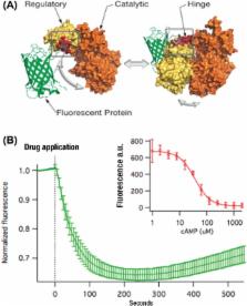
          Protein-based, fluorescent biosensors power basic research on cell signaling in health and disease, but their use in automated laboratories is limited. We have now created two live-cell assays, one for diacyl glycerol and another for cAMP, that are robust (Z′ > 0.7) and easily deployed on standard fluorescence plate readers. We describe the development of these assays, focusing on the parameters that were critical for optimization, in the hopes that the lessons learned can be generalized to the development of new biosensor-based assays.

          Related collections

          Most cited references24

          • Record: found
          • Abstract: found
          • Article: not found

          A bright monomeric green fluorescent protein derived from Branchiostoma lanceolatum

          Despite the existence of fluorescent proteins spanning the entire visual spectrum, the bulk of modern imaging experiments continue to rely on variants of the green fluorescent protein derived from Aequorea victoria. Meanwhile, a great deal of recent effort has been devoted to engineering and improving red fluorescent proteins, and relatively little attention has been given to green and yellow variants. Here we report a novel monomeric yellow-green fluorescent protein, mNeonGreen, which is derived from a tetrameric fluorescent protein from the cephalochordate Branchiostoma lanceolatum. This fluorescent protein is the brightest monomeric green or yellow fluorescent protein yet described, performs exceptionally well as a fusion tag for traditional imaging as well as stochastic single-molecule superresolution imaging, and is an excellent FRET acceptor for the newest generation of cyan fluorescent proteins.
            Bookmark
            • Record: found
            • Abstract: found
            • Article: not found

            Optimization of a GCaMP calcium indicator for neural activity imaging.

            Genetically encoded calcium indicators (GECIs) are powerful tools for systems neuroscience. Recent efforts in protein engineering have significantly increased the performance of GECIs. The state-of-the art single-wavelength GECI, GCaMP3, has been deployed in a number of model organisms and can reliably detect three or more action potentials in short bursts in several systems in vivo. Through protein structure determination, targeted mutagenesis, high-throughput screening, and a battery of in vitro assays, we have increased the dynamic range of GCaMP3 by severalfold, creating a family of "GCaMP5" sensors. We tested GCaMP5s in several systems: cultured neurons and astrocytes, mouse retina, and in vivo in Caenorhabditis chemosensory neurons, Drosophila larval neuromuscular junction and adult antennal lobe, zebrafish retina and tectum, and mouse visual cortex. Signal-to-noise ratio was improved by at least 2- to 3-fold. In the visual cortex, two GCaMP5 variants detected twice as many visual stimulus-responsive cells as GCaMP3. By combining in vivo imaging with electrophysiology we show that GCaMP5 fluorescence provides a more reliable measure of neuronal activity than its predecessor GCaMP3. GCaMP5 allows more sensitive detection of neural activity in vivo and may find widespread applications for cellular imaging in general.
              Bookmark
              • Record: found
              • Abstract: found
              • Article: not found

              An optimized fluorescent probe for visualizing glutamate neurotransmission

              We describe an intensity-based glutamate-sensing fluorescent reporter (“iGluSnFR”) with signal-to-noise ratio and kinetics appropriate for in vivo imaging. We engineered iGluSnFR in vitro to maximize its fluorescence change, and validated its utility for visualizing glutamate release by neurons and astrocytes in increasingly intact neurological systems. In hippocampal culture, iGluSnFR detected single field stimulus-evoked glutamate release events. In pyramidal neurons in acute brain slices, glutamate uncaging at single spines showed that iGluSnFR responds robustly and specifically to glutamate in situ, and responses correlate with voltage changes. In mouse retina, iGluSnFR-expressing neurons showed intact light-evoked excitatory currents, and the sensor revealed tonic glutamate signaling in response to light stimuli. In worms, glutamate signals preceded and predicted post-synaptic calcium transients. In zebrafish, iGluSnFR revealed spatial organization of direction-selective synaptic activity in the optic tectum. Finally, in mouse forelimb motor cortex, iGluSnFR expression in layer V pyramidal neurons revealed task-dependent single-spine activity during running.
                Bookmark

                Author and article information

                Journal
                J Biomol Screen
                J Biomol Screen
                JBX
                spjbx
                Journal of Biomolecular Screening
                SAGE Publications (Sage CA: Los Angeles, CA )
                1087-0571
                1552-454X
                11 December 2015
                March 2016
                : 21
                : 3
                : 298-305
                Affiliations
                [1 ]Montana Molecular, Bozeman, MT, USA
                [2 ]Scintillon Institute, San Diego, CA, USA
                Author notes
                [*]Anne Marie Quinn, Montana Molecular, 347 S. Ferguson Avenue, Bozeman, MT 59718, USA. Email: amq@ 123456montanamolecular.com
                Article
                10.1177_1087057115618608
                10.1177/1087057115618608
                4766961
                26657040
                e2578ad6-53b4-4ce3-90ee-f2077e6fc934
                © 2015 Society for Laboratory Automation and Screening

                This article is distributed under the terms of the Creative Commons Attribution-NonCommercial 3.0 License ( http://www.creativecommons.org/licenses/by-nc/3.0/) which permits non-commercial use, reproduction and distribution of the work without further permission provided the original work is attributed as specified on the SAGE and Open Access page ( https://us.sagepub.com/en-us/nam/open-access-at-sage).

                History
                : 7 August 2015
                : 1 October 2015
                : 13 October 2015
                Categories
                Original Research

                Molecular medicine
                fluorescence,baculovirus,mneongreen,gfp,z′ statistic,live-cell assay,adenylyl cyclase,camp,diacyl glycerol,dag

                Comments

                Comment on this article