6
views
0
recommends
+1 Recommend
0 collections
    0
    shares
      • Record: found
      • Abstract: found
      • Article: found
      Is Open Access

      The Role of Innate Leukocytes during Influenza Virus Infection

      review-article
      1 , 2 , 1 , 2 ,
      Journal of Immunology Research
      Hindawi

      Read this article at

      Bookmark
          There is no author summary for this article yet. Authors can add summaries to their articles on ScienceOpen to make them more accessible to a non-specialist audience.

          Abstract

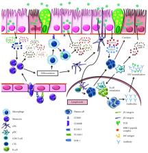
          Influenza virus infection is a serious threat to humans and animals, with the potential to cause severe pneumonia and death. Annual vaccination strategies are a mainstay to prevent complications related to influenza. However, protection from the emerging subtypes of influenza A viruses (IAV) even in vaccinated individuals is challenging. Innate immune cells are the first cells to respond to IAV infection in the respiratory tract. Virus replication-induced production of cytokines from airway epithelium recruits innate immune cells to the site of infection. These leukocytes, namely, neutrophils, monocytes, macrophages, dendritic cells, eosinophils, natural killer cells, innate lymphoid cells, and γδ T cells, become activated in response to IAV, to contain the virus and protect the airway epithelium while triggering the adaptive arm of the immune system. This review addresses different anti-influenza virus schemes of innate immune cells and how these cells fine-tune the balance between immunoprotection and immunopathology during IAV infection. Detailed understanding on how these innate responders execute anti-influenza activity will help to identify novel therapeutic targets to halt IAV replication and associated immunopathology.

          Related collections

          Most cited references141

          • Record: found
          • Abstract: found
          • Article: not found

          Alveolar macrophages: plasticity in a tissue-specific context.

          Alveolar macrophages exist in a unique microenvironment and, despite historical evidence showing that they are in close contact with the respiratory epithelium, have until recently been investigated in isolation. The microenvironment of the airway lumen has a considerable influence on many aspects of alveolar macrophage phenotype, function and turnover. As the lungs adapt to environmental challenges, so too do alveolar macrophages adapt to accommodate the ever-changing needs of the tissue. In this Review, we discuss the unique characteristics of alveolar macrophages, the mechanisms that drive their adaptation and the direct and indirect influences of epithelial cells on them. We also highlight how airway luminal macrophages function as sentinels of a healthy state and how they do not respond in a pro-inflammatory manner to antigens that do not disrupt lung structure. The unique tissue location and function of alveolar macrophages distinguish them from other macrophage populations and suggest that it is important to classify macrophages according to the site that they occupy.
            Bookmark
            • Record: found
            • Abstract: found
            • Article: not found

            Excessive Neutrophils and Neutrophil Extracellular Traps Contribute to Acute Lung Injury of Influenza Pneumonitis

            Complications of acute respiratory distress syndrome (ARDS) are common among critically ill patients infected with highly pathogenic influenza viruses. Macrophages and neutrophils constitute the majority of cells recruited into infected lungs, and are associated with immunopathology in influenza pneumonia. We examined pathological manifestations in models of macrophage- or neutrophil-depleted mice challenged with sublethal doses of influenza A virus H1N1 strain PR8. Infected mice depleted of macrophages displayed excessive neutrophilic infiltration, alveolar damage, and increased viral load, later progressing into ARDS-like pathological signs with diffuse alveolar damage, pulmonary edema, hemorrhage, and hypoxemia. In contrast, neutrophil-depleted animals showed mild pathology in lungs. The brochoalveolar lavage fluid of infected macrophage-depleted mice exhibited elevated protein content, T1-α, thrombomodulin, matrix metalloproteinase-9, and myeloperoxidase activities indicating augmented alveolar-capillary damage, compared to neutrophil-depleted animals. We provide evidence for the formation of neutrophil extracellular traps (NETs), entangled with alveoli in areas of tissue injury, suggesting their potential link with lung damage. When co-incubated with infected alveolar epithelial cells in vitro, neutrophils from infected lungs strongly induced NETs generation, and augmented endothelial damage. NETs induction was abrogated by anti-myeloperoxidase antibody and an inhibitor of superoxide dismutase, thus implying that NETs generation is induced by redox enzymes in influenza pneumonia. These findings support the pathogenic effects of excessive neutrophils in acute lung injury of influenza pneumonia by instigating alveolar-capillary damage.
              Bookmark
              • Record: found
              • Abstract: found
              • Article: not found

              NETosis: how vital is it?

              In this review, we examine the evidence that neutrophil extracellular traps (NETs) play a critical role in innate immunity. We summarize how NETs are formed in response to various stimuli and provide evidence that NETosis is not universally a cell death pathway. Here we describe at least 2 different mechanisms by which NETs are formed, including a suicide lytic NETosis and a live cell or vital NETosis. We also evaluate the evidence for NETs in catching and killing pathogens. Finally, we examine how infections are related to the development of autoimmune and vasculitic diseases through unintended but detrimental bystander damage resulting from NET release.
                Bookmark

                Author and article information

                Contributors
                Journal
                J Immunol Res
                J Immunol Res
                JIR
                Journal of Immunology Research
                Hindawi
                2314-8861
                2314-7156
                2019
                12 September 2019
                : 2019
                : 8028725
                Affiliations
                1Department of Pediatrics, University of Tennessee Health Science Center, Memphis, TN 38103, USA
                2Children's Foundation Research Institute, Memphis, TN 38103, USA
                Author notes

                Guest Editor: Patricio L. Acosta

                Author information
                https://orcid.org/0000-0002-3104-2823
                Article
                10.1155/2019/8028725
                6757286
                31612153
                e28ab873-275e-4dfb-99fb-8e763d26a14c
                Copyright © 2019 Prem P. Lamichhane and Amali E. Samarasinghe.

                This is an open access article distributed under the Creative Commons Attribution License, which permits unrestricted use, distribution, and reproduction in any medium, provided the original work is properly cited.

                History
                : 9 April 2019
                : 15 July 2019
                Funding
                Funded by: National Institutes of Health
                Award ID: R01-AI125481
                Categories
                Review Article

                Comments

                Comment on this article