17
views
0
recommends
+1 Recommend
0 collections
    0
    shares
      • Record: found
      • Abstract: found
      • Article: found
      Is Open Access

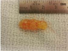
      Papillary fibroelastoma arising from the coumadin ridge

      case-report

      Read this article at

      Bookmark
          There is no author summary for this article yet. Authors can add summaries to their articles on ScienceOpen to make them more accessible to a non-specialist audience.

          Abstract

          Cardiac papillary fibroelastomas (CPF) are rare cardiac tumors, mostly found on the valvular surfaces in the heart. These tumors are frond like in nature and are benign, intracardiac masses, rarely causing any hemodynamic disturbances. However, excision of these masses is indicated due to their propensity to embolize. We present a case report of the tumor found on the coumadin ridge, causing transient ischemic attacks in a patient. We performed complete excision of the tumor via median sternotomy on cardiopulmonary bypass support with cardiac arrest. The diagnosis was confirmed by histological examination. The patient had an uneventful postoperative course and was discharghed on postoperative day 4. She has had complete resolution of her symptoms post excision. The diagnosis of the mass was confirmed on histological examination.

          Related collections

          Most cited references8

          • Record: found
          • Abstract: found
          • Article: not found

          Cardiac papillary fibroelastoma: a comprehensive analysis of 725 cases.

          With the advent of echocardiography, cardiac papillary fibroelastoma (CPF) is being increasingly reported. The demographics, clinical characteristics, pathological features, treatment, and prognosis of CPF are examined. Cases, case series and related articles on the subject in all languages were identified through a comprehensive literature search. Seven hundred twenty-five cases of CPF were identified. Males comprised 55% of patients. Highest prevalence was in the 8th decade of life. The valvular surface was the predominant locations of tumor. The most commonly involved valve was the aortic valve, followed by the mitral valve. The left ventricle was the predominant nonvalvular site involved. No clear risk factor for development of CPF has been reported. Size of the tumor varied from 2 mm to 70 mm. Clinically, CPFs have presented with transient ischemic attack, stroke, myocardial infarction, sudden death, heart failure, presyncope, syncope, pulmonary embolism, blindness, and peripheral embolism. Tumor mobility was the only independent predictor of CPF-related death or nonfatal embolization. Symptomatic patients should be treated surgically because the successful complete resection of CPF is curative and the long-term postoperative prognosis is excellent. The symptomatic patients who are not surgical candidates could be offered long-term oral anticoagulation, although no randomized controlled data are available on its efficacy. Asymptomatic patients could be treated surgically if the tumor is mobile, as the tumor mobility is the independent predictor of death or nonfatal embolization. Asymptomatic patients with nonmobile CPF could be followed-up closely with periodic clinical evaluation and echocardiography, and receive surgical intervention when symptoms develop or the tumor becomes mobile.
            Bookmark
            • Record: found
            • Abstract: found
            • Article: not found

            Primary and metastatic cardiac tumors: imaging characteristics, surgical treatment, and histopathological spectrum: a 10-year-experience at a German heart center.

            Tumors of the heart are rare compared to other cardiac diseases. Their clinical symptoms vary from absent to nonspecific. This great variation and general paucity of symptoms related to cardiac tumors often result in delayed diagnosis and treatment. We retrospectively evaluated all patients who underwent cardiac surgery for a space-occupying lesion in the observation period between 2000 and 2010 at our hospital. Clinicopathological features, imaging characteristics, and disease outcomes were analyzed, and the results were compared with the available English literature. During the last 10 years, 84 patients underwent resection of a cardiac mass at our center, i.e., 0.85% of the total number (n=9829) of all cardiac surgical operations performed in that period. The part of primary cardiac tumors was 73.8% (n=62; 59 benign and 3 malign tumors). In nine cases (10.7%), secondary cardiac tumors represented metastases of malignant tumors from different extracardiac locations. In 13 cases (15.5%), the mass represented cardiac thrombus. The majority of cardiac tumors were benign, and most of them were cardiac myxomas (n=48). Papillary fibroelastoma was the second most common primary tumor. Our data in this study were comparable to the literature regarding the frequency and allocation of the different cardiac tumors. Our data are in line with previous reports that patients with benign cardiac tumors profit from surgical resection compared to those subjected to conservative treatment with the risk of central or peripheral embolisms. Concerning malignant primary cardiac tumors and cardiac metastases, surgery represents only a palliative strategy in most of the cases. Copyright © 2012 Elsevier Inc. All rights reserved.
              Bookmark
              • Record: found
              • Abstract: not found
              • Article: not found

              Role of magnetic resonance imaging for evaluation of tumors in the cardiac region

                Bookmark

                Author and article information

                Journal
                J Cardiovasc Thorac Res
                J Cardiovasc Thorac Res
                J Cardiovasc Thorac Res
                JCVTR
                TBZMED
                Journal of Cardiovascular and Thoracic Research
                Tabriz University of Medical Sciences
                2008-5117
                2008-6830
                2017
                12 April 2017
                : 9
                : 2
                : 118-120
                Affiliations
                1Division of Thoracic Surgery, Department of Surgery, The Ohio State University Wexner Medical Center, Columbus, OH, USA
                2Department of Pathology, The Ohio State University Wexner Medical Center, Columbus, OH, USA
                Author notes
                [* ] Corresponding Author: Mahim Malik, Email: mahim.malik@ 123456osumc.edu
                Article
                10.15171/jcvtr.2017.20
                5516052
                e32f4de6-f4c6-45fa-9e93-3d7a5f1c581c
                © 2017 The Author(s)

                This is an open access article distributed under the terms of the Creative Commons Attribution License ( http://creativecommons.org/licenses/by/4.0/), which permits unrestricted use, distribution, and reproduction in any medium, provided the original work is properly cited.

                History
                : 02 December 2015
                : 29 August 2016
                Page count
                Figures: 4, References: 9, Pages: 3
                Categories
                Case Report

                fibroelastoma,tumor,left atrium
                fibroelastoma, tumor, left atrium

                Comments

                Comment on this article